Cytokeratin 10 Polyclonal Antibody
- Catalog No.:YT1259
- Applications:WB;IHC;IF;ELISA
- Reactivity:Human;Mouse;Rat
- Target:
- Cytokeratin 10
- Fields:
- >>Estrogen signaling pathway;>>Staphylococcus aureus infection
- Gene Name:
- KRT10
- Protein Name:
- Keratin type I cytoskeletal 10
- Human Gene Id:
- 3858
- Human Swiss Prot No:
- P13645
- Mouse Gene Id:
- 16661
- Mouse Swiss Prot No:
- P02535
- Rat Gene Id:
- 450225
- Rat Swiss Prot No:
- Q6IFW6
- Immunogen:
- The antiserum was produced against synthesized peptide derived from human Keratin 10. AA range:136-185
- Specificity:
- Cytokeratin 10 Polyclonal Antibody detects endogenous levels of Cytokeratin 10 protein.
- Formulation:
- Liquid in PBS containing 50% glycerol, 0.5% BSA and 0.02% sodium azide.
- Source:
- Polyclonal, Rabbit,IgG
- Dilution:
- WB 1:500 - 1:2000. IHC 1:100 - 1:300. IF 1:200 - 1:1000. ELISA: 1:20000. Not yet tested in other applications.
- Purification:
- The antibody was affinity-purified from rabbit antiserum by affinity-chromatography using epitope-specific immunogen.
- Concentration:
- 1 mg/ml
- Storage Stability:
- -15°C to -25°C/1 year(Do not lower than -25°C)
- Other Name:
- KRT10;KPP;Keratin; type I cytoskeletal 10;Cytokeratin-10;CK-10;Keratin-10;K10
- Observed Band(KD):
- 59kD
- Background:
- This gene encodes a member of the type I (acidic) cytokeratin family, which belongs to the superfamily of intermediate filament (IF) proteins. Keratins are heteropolymeric structural proteins which form the intermediate filament. These filaments, along with actin microfilaments and microtubules, compose the cytoskeleton of epithelial cells. Mutations in this gene are associated with epidermolytic hyperkeratosis. This gene is located within a cluster of keratin family members on chromosome 17q21. [provided by RefSeq, Jul 2008],
- Function:
- disease:Defects in KRT10 are a cause of bullous congenital ichthyosiform erythroderma (BCIE) [MIM:113800]; also known as epidermolytic hyperkeratosis (EHK) or bullous erythroderma ichthyosiformis congenita of Brocq. BCIE is an autosomal dominant skin disorder characterized by widespread blistering and an ichthyotic erythroderma at birth that persist into adulthood. Histologically there is a diffuse epidermolytic degeneration in the lower spinous layer of the epidermis. Within a few weeks from birth, erythroderma and blister formation diminish and hyperkeratoses develop.,disease:Defects in KRT10 are a cause of epidermal nevus epidermolytic hyperkeratotic type [MIM:600648]. Epidermal nevi affect about 1 in 1,000 people. They appear at or shortly after birth as localized lines of epidermal thickening. The extent of skin involvement varies widely.,disease:Defects in KRT10 are a cause of icht
- Subcellular Location:
- Secreted, extracellular space . Cell surface . Localized on the surface of desquamated nasal epithelial cells (PubMed:12427098). Localized on the surface of lung cell lines (PubMed:19627498). .
- Expression:
- Seen in all suprabasal cell layers including stratum corneum. Expressed on the surface of lung cell lines (PubMed:19627498).
- June 19-2018
- WESTERN IMMUNOBLOTTING PROTOCOL
- June 19-2018
- IMMUNOHISTOCHEMISTRY-PARAFFIN PROTOCOL
- June 19-2018
- IMMUNOFLUORESCENCE PROTOCOL
- September 08-2020
- FLOW-CYTOMEYRT-PROTOCOL
- May 20-2022
- Cell-Based ELISA│解您多样本WB检测之困扰
- July 13-2018
- CELL-BASED-ELISA-PROTOCOL-FOR-ACETYL-PROTEIN
- July 13-2018
- CELL-BASED-ELISA-PROTOCOL-FOR-PHOSPHO-PROTEIN
- July 13-2018
- Antibody-FAQs
- Products Images
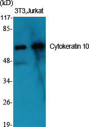
- Western Blot analysis of various cells using Cytokeratin 10 Polyclonal Antibody
.jpg)
- Western Blot analysis of HeLa cells using Cytokeratin 10 Polyclonal Antibody
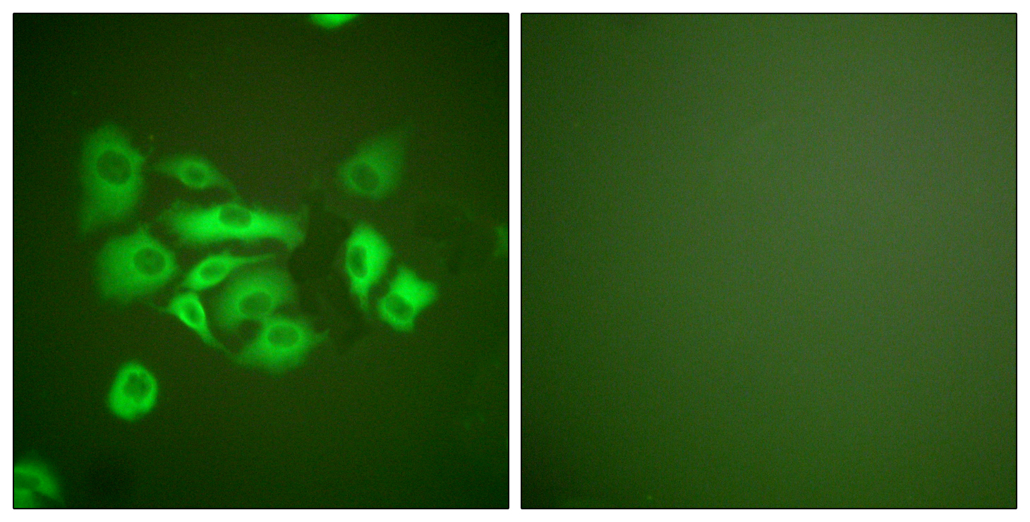
- Immunofluorescence analysis of A549 cells, using Keratin 10 Antibody. The picture on the right is blocked with the synthesized peptide.
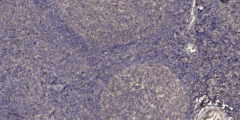
- Immunohistochemical analysis of paraffin-embedded human tonsil. 1, Antibody was diluted at 1:200(4° overnight). 2, Tris-EDTA,pH9.0 was used for antigen retrieval. 3,Secondary antibody was diluted at 1:200(room temperature, 30min).


