c-Myc Polyclonal Antibody
- Catalog No.:YT0991
- Applications:WB;IHC;IF;ELISA
- Reactivity:Human;Mouse;Rat
- Target:
- c-Myc
- Fields:
- >>MAPK signaling pathway;>>ErbB signaling pathway;>>Cell cycle;>>PI3K-Akt signaling pathway;>>Cellular senescence;>>Wnt signaling pathway;>>TGF-beta signaling pathway;>>Hippo signaling pathway;>>Signaling pathways regulating pluripotency of stem cells;>>JAK-STAT signaling pathway;>>Thyroid hormone signaling pathway;>>Salmonella infection;>>Hepatitis C;>>Hepatitis B;>>Human cytomegalovirus infection;>>Human T-cell leukemia virus 1 infection;>>Kaposi sarcoma-associated herpesvirus infection;>>Epstein-Barr virus infection;>>Pathways in cancer;>>Transcriptional misregulation in cancer;>>Proteoglycans in cancer;>>MicroRNAs in cancer;>>Chemical carcinogenesis - receptor activation;>>Colorectal cancer;>>Endometrial cancer;>>Thyroid cancer;>>Bladder cancer;>>Chronic myeloid leukemia;>>Acute myeloid leukemia;>>Small cell lung cancer;>>Breast cancer;>>Hepatocellular carcinoma;>>Gastric cancer;>>Central carbon metabolism in cancer
- Gene Name:
- MYC
- Protein Name:
- Myc proto-oncogene protein
- Human Gene Id:
- 4609
- Human Swiss Prot No:
- P01106
- Mouse Gene Id:
- 17869
- Mouse Swiss Prot No:
- P01108
- Rat Gene Id:
- 24577
- Rat Swiss Prot No:
- P09416
- Immunogen:
- The antiserum was produced against synthesized peptide derived from human MYC. AA range:386-435
- Specificity:
- c-Myc Polyclonal Antibody detects endogenous levels of c-Myc protein.
- Formulation:
- Liquid in PBS containing 50% glycerol, 0.5% BSA and 0.02% sodium azide.
- Source:
- Polyclonal, Rabbit,IgG
- Dilution:
- WB 1:500 - 1:2000. IHC 1:100 - 1:300. IF 1:200 - 1:1000. ELISA: 1:40000. Not yet tested in other applications.
- Purification:
- The antibody was affinity-purified from rabbit antiserum by affinity-chromatography using epitope-specific immunogen.
- Concentration:
- 1 mg/ml
- Storage Stability:
- -15°C to -25°C/1 year(Do not lower than -25°C)
- Other Name:
- MYC;BHLHE39;Myc proto-oncogene protein;Class E basic helix-loop-helix protein 39;bHLHe39;Proto-oncogene c-Myc;Transcription factor p64
- Observed Band(KD):
- 50,(also ~60KD in some samples)
- Background:
- The protein encoded by this gene is a multifunctional, nuclear phosphoprotein that plays a role in cell cycle progression, apoptosis and cellular transformation. It functions as a transcription factor that regulates transcription of specific target genes. Mutations, overexpression, rearrangement and translocation of this gene have been associated with a variety of hematopoietic tumors, leukemias and lymphomas, including Burkitt lymphoma. There is evidence to show that alternative translation initiations from an upstream, in-frame non-AUG (CUG) and a downstream AUG start site result in the production of two isoforms with distinct N-termini. The synthesis of non-AUG initiated protein is suppressed in Burkitt's lymphomas, suggesting its importance in the normal function of this gene. [provided by RefSeq, Jul 2008],
- Function:
- disease:A chromosomal aberration involving MYC may be a cause of a form of B-cell chronic lymphocytic leukemia. Translocation t(8;12)(q24;q22) with BTG1.,disease:Overexpression of MYC is implicated in the etiology of a variety of hematopoietic tumors.,function:Participates in the regulation of gene transcription. Binds DNA both in a non-specific manner and also specifically to recognizes the core sequence 5'-CAC[GA]TG-3'. Seems to activate the transcription of growth-related genes.,online information:Myc entry,PTM:Phosphorylated by PRKDC.,similarity:Contains 1 basic helix-loop-helix (bHLH) domain.,subunit:Efficient DNA binding requires dimerization with another bHLH protein. Binds DNA as a heterodimer with MAX. Interacts with TAF1C and SPAG9. Interacts with PARP10. Interacts with KDM5A and KDM5B.,
- Subcellular Location:
- Nucleus, nucleoplasm . Nucleus, nucleolus .
- Expression:
- Cervix,Epithelium,Leukemia,Placenta,Promyelocytic l
DNA damage to bone marrow stromal cells by anti-leukemia drugs induces chemo-resistance in acute myeloid leukemia via paracrine FGF10-FGFR2 signaling JOURNAL OF BIOLOGICAL CHEMISTRY Shuang Yu, Jingjing Ye, Yingqiao Wang, Ting Lu, Yan Liu, Na Liu, Jingru Zhang, Fei Lu, Daoxin Ma, Robert Peter Gale, Chunyan Ji WB Human HS-5 cell
Licoflavone A Suppresses Gastric Cancer Growth and Metastasis by Blocking the VEGFR-2 Signaling Pathway. Journal of Oncology2022;2022:5497991. Human 1 : 1000 MKN-45 cell
HMGA1-TRIP13 axis promotes stemness and epithelial mesenchymal transition of perihilar cholangiocarcinoma in a positive feedback loop dependent on c-Myc. JOURNAL OF EXPERIMENTAL & CLINICAL CANCER RESEARCH J Exp Clin Canc Res. 2021 Dec;40(1):1-18 WB Human pCCA tissues ,adjacent normal tissue QBC939 cell,FRH0201 cell
Hepatomas are exquisitely sensitive to pharmacologic ascorbate (P-AscH-). Theranostics Theranostics. 2019; 9(26): 8109–8126 WB Human Huh-7 cell
Identification of WISP1 as a novel oncogene in glioblastoma. INTERNATIONAL JOURNAL OF ONCOLOGY Int J Oncol. 2017 Oct;51(4):1261-1270 WB Human 1:1000 U251 cell, U373-MG cell
In Utero Exposure to Fine Particulate Matter Causes Hypertension Due to Impaired Renal Dopamine D1 Receptor in Offspring. CELLULAR PHYSIOLOGY AND BIOCHEMISTRY Cell Physiol Biochem. 2018;46(1):148-159 WB Rat 1:500 renal cortex
Resveratrol Ameliorates the Anxiety- and Depression-Like Behavior of Subclinical Hypothyroidism Rat: Possible Involvement of the HPT Axis, HPA Axis, and Wnt/β-Catenin Pathway. Frontiers in Endocrinology Front Endocrinol. 2016 May;0:44 WB Rat 1:1000 hippocampus
Role of Notch pathway in effect of mono-2-ethylhexyl phthalate on the proliferation and cell cycle of SH-SY5Y cell. ENVIRONMENTAL TOXICOLOGY Environ Toxicol. 2021 Sep;36(9):1944-1952 WB Human SH-SY5Y cell
Chronic CagA‐positive Helicobacter pylori infection with MNNG stimulation synergistically induces mesenchymal and cancer stem cell‐like properties in gastric mucosal epithelial cells. JOURNAL OF CELLULAR BIOCHEMISTRY J Cell Biochem. 2019 Oct;120(10):17635-17649 WB Human GES-1 cell
S100A4 promotes colon inflammation and colitis-associated colon tumorigenesis. OncoImmunology 2018 Jun 11 WB Mouse colon tissues macrophage
Maekawa, Fumihiko. "Jin-Fang Ge*†, Ya-Yun Xu†, Gan Qin, Jiang-Qun Cheng and Fei-Hu Chen." Chemicals in the Environment and Brain Development: Importance of Neuroendocrinological Approaches (2017): 138.
Design, synthesis and biological evaluation of coumarin derivatives as potential BRD4 inhibitors BIOORGANIC CHEMISTRY Shi-Wu Chen WB Human
- June 19-2018
- WESTERN IMMUNOBLOTTING PROTOCOL
- June 19-2018
- IMMUNOHISTOCHEMISTRY-PARAFFIN PROTOCOL
- June 19-2018
- IMMUNOFLUORESCENCE PROTOCOL
- September 08-2020
- FLOW-CYTOMEYRT-PROTOCOL
- May 20-2022
- Cell-Based ELISA│解您多样本WB检测之困扰
- July 13-2018
- CELL-BASED-ELISA-PROTOCOL-FOR-ACETYL-PROTEIN
- July 13-2018
- CELL-BASED-ELISA-PROTOCOL-FOR-PHOSPHO-PROTEIN
- July 13-2018
- Antibody-FAQs
- Products Images
- Ye, Zhengmeng, et al. "In Utero Exposure to Fine Particulate Matter Causes Hypertension Due to Impaired Renal Dopamine D1 Receptor in Offspring." Cellular Physiology and Biochemistry 46.1 (2018): 148-159.
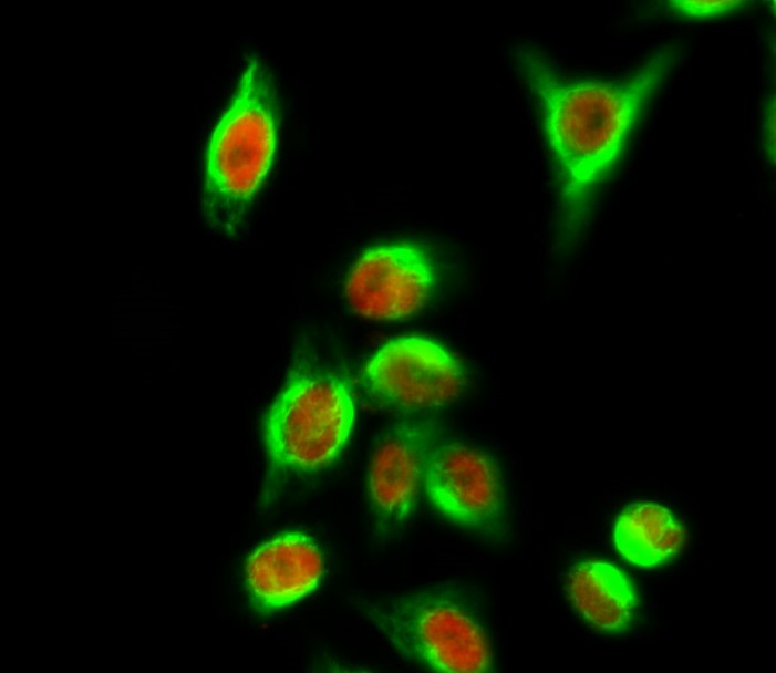
- Immunofluorescence analysis of Hela cell. 1,c-Myc Polyclonal Antibody(red) was diluted at 1:200(4° overnight). CYCS Monoclonal Antibody(4B10)(green) was diluted at 1:200(4° overnight). 2, Goat Anti Rabbit Alexa Fluor 594 Catalog:RS3611 was diluted at 1:1000(room temperature, 50min). Goat Anti Mouse Alexa Fluor 488 Catalog:RS3208 was diluted at 1:1000(room temperature, 50min).
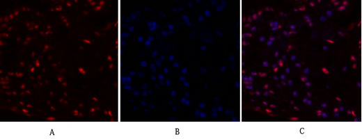
- Immunofluorescence analysis of human-lung tissue. 1,c-Myc Polyclonal Antibody(red) was diluted at 1:200(4°C,overnight). 2, Cy3 labled Secondary antibody was diluted at 1:300(room temperature, 50min).3, Picture B: DAPI(blue) 10min. Picture A:Target. Picture B: DAPI. Picture C: merge of A+B
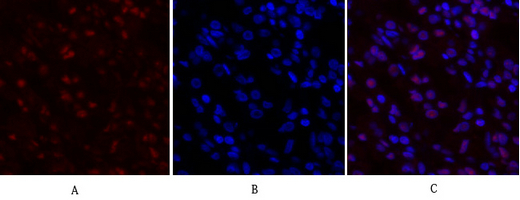
- Immunofluorescence analysis of human-stomach tissue. 1,c-Myc Polyclonal Antibody(red) was diluted at 1:200(4°C,overnight). 2, Cy3 labled Secondary antibody was diluted at 1:300(room temperature, 50min).3, Picture B: DAPI(blue) 10min. Picture A:Target. Picture B: DAPI. Picture C: merge of A+B
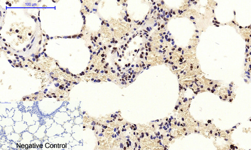
- Immunohistochemical analysis of paraffin-embedded Rat-lung tissue. 1,c-Myc Polyclonal Antibody was diluted at 1:200(4°C,overnight). 2, Sodium citrate pH 6.0 was used for antibody retrieval(>98°C,20min). 3,Secondary antibody was diluted at 1:200(room tempeRature, 30min). Negative control was used by secondary antibody only.

- Immunohistochemical analysis of paraffin-embedded Rat-brain tissue. 1,c-Myc Polyclonal Antibody was diluted at 1:200(4°C,overnight). 2, Sodium citrate pH 6.0 was used for antibody retrieval(>98°C,20min). 3,Secondary antibody was diluted at 1:200(room tempeRature, 30min). Negative control was used by secondary antibody only.
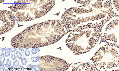
- Immunohistochemical analysis of paraffin-embedded Mouse-testis tissue. 1,c-Myc Polyclonal Antibody was diluted at 1:200(4°C,overnight). 2, Sodium citrate pH 6.0 was used for antibody retrieval(>98°C,20min). 3,Secondary antibody was diluted at 1:200(room tempeRature, 30min). Negative control was used by secondary antibody only.
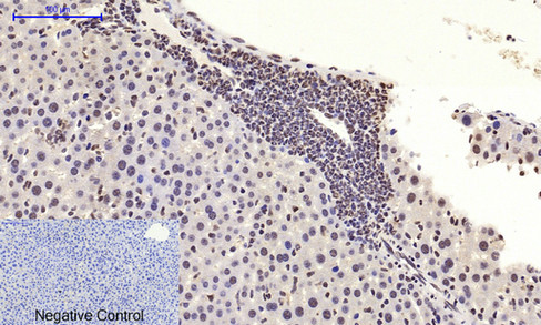
- Immunohistochemical analysis of paraffin-embedded Mouse-liver tissue. 1,c-Myc Polyclonal Antibody was diluted at 1:200(4°C,overnight). 2, Sodium citrate pH 6.0 was used for antibody retrieval(>98°C,20min). 3,Secondary antibody was diluted at 1:200(room tempeRature, 30min). Negative control was used by secondary antibody only.
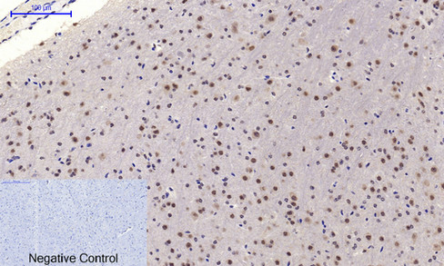
- Immunohistochemical analysis of paraffin-embedded Mouse-brain tissue. 1,c-Myc Polyclonal Antibody was diluted at 1:200(4°C,overnight). 2, Sodium citrate pH 6.0 was used for antibody retrieval(>98°C,20min). 3,Secondary antibody was diluted at 1:200(room tempeRature, 30min). Negative control was used by secondary antibody only.
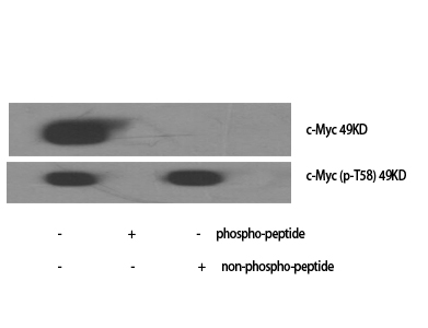
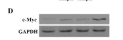
- Western Blot analysis of various cells using c-Myc Polyclonal Antibody diluted at 1:1000
.jpg)
- Western Blot analysis of HuvEc cells using c-Myc Polyclonal Antibody diluted at 1:1000
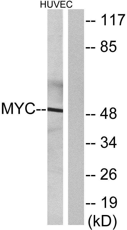
- Western blot analysis of lysates from HUVEC cells, using MYC Antibody. The lane on the right is blocked with the synthesized peptide.


