ARHGAP9 Polyclonal Antibody
- Catalog No.:YT0320
- Applications:WB;IHC;IF;ELISA
- Reactivity:Human;Rat;Mouse;
- Target:
- ARHGAP9
- Gene Name:
- ARHGAP9
- Protein Name:
- Rho GTPase-activating protein 9
- Human Gene Id:
- 64333
- Human Swiss Prot No:
- Q9BRR9
- Immunogen:
- The antiserum was produced against synthesized peptide derived from human RHG9. AA range:220-269
- Specificity:
- ARHGAP9 Polyclonal Antibody detects endogenous levels of ARHGAP9 protein.
- Formulation:
- Liquid in PBS containing 50% glycerol, 0.5% BSA and 0.02% sodium azide.
- Source:
- Polyclonal, Rabbit,IgG
- Dilution:
- WB 1:500 - 1:2000. IHC 1:100 - 1:300. ELISA: 1:40000.. IF 1:50-200
- Purification:
- The antibody was affinity-purified from rabbit antiserum by affinity-chromatography using epitope-specific immunogen.
- Concentration:
- 1 mg/ml
- Storage Stability:
- -15°C to -25°C/1 year(Do not lower than -25°C)
- Other Name:
- ARHGAP9;Rho GTPase-activating protein 9;Rho-type GTPase-activating protein 9
- Observed Band(KD):
- 90kD
- Background:
- This gene encodes a member of the Rho-GAP family of GTPase activating proteins. The protein has substantial GAP activity towards several Rho-family GTPases in vitro, converting them to an inactive GDP-bound state. It is implicated in regulating adhesion of hematopoietic cells to the extracellular matrix. Multiple transcript variants encoding different isoforms have been found for this gene. [provided by RefSeq, Jul 2008],
- Function:
- function:GTPase activator for the Rho-type GTPases by converting them to an inactive GDP-bound state. Has a substantial GAP activity toward CDC42 and RAC1 and less toward RHOA. Has a role in regulating adhesion of hematopoietic cells to the extracellular matrix.,similarity:Contains 1 PH domain.,similarity:Contains 1 Rho-GAP domain.,similarity:Contains 1 SH3 domain.,similarity:Contains 1 WW domain.,tissue specificity:Predominantly expressed in peripheral blood leukocytes, spleen, and thymus.,
- Subcellular Location:
- cytosol,
- Expression:
- Predominantly expressed in peripheral blood leukocytes, spleen, and thymus.
- June 19-2018
- WESTERN IMMUNOBLOTTING PROTOCOL
- June 19-2018
- IMMUNOHISTOCHEMISTRY-PARAFFIN PROTOCOL
- June 19-2018
- IMMUNOFLUORESCENCE PROTOCOL
- September 08-2020
- FLOW-CYTOMEYRT-PROTOCOL
- May 20-2022
- Cell-Based ELISA│解您多样本WB检测之困扰
- July 13-2018
- CELL-BASED-ELISA-PROTOCOL-FOR-ACETYL-PROTEIN
- July 13-2018
- CELL-BASED-ELISA-PROTOCOL-FOR-PHOSPHO-PROTEIN
- July 13-2018
- Antibody-FAQs
- Products Images
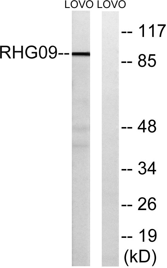
- Western blot analysis of lysates from LOVO cells, using RHG9 Antibody. The lane on the right is blocked with the synthesized peptide.
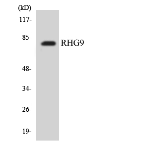
- Western blot analysis of the lysates from COLO205 cells using RHG9 antibody.

- Immunohistochemical analysis of paraffin-embedded human Gastric adenocarcinoma. 1, Antibody was diluted at 1:200(4° overnight). 2, Tris-EDTA,pH9.0 was used for antigen retrieval. 3,Secondary antibody was diluted at 1:200(room temperature, 45min).


