AMPKα1/2 Polyclonal Antibody
- Catalog No.:YT0216
- Applications:WB;IHC;IF;ELISA
- Reactivity:Human;Mouse;Rat;Monkey;Bovine;Fish
- Target:
- AMPKα1/2
- Fields:
- >>FoxO signaling pathway;>>Autophagy - animal;>>mTOR signaling pathway;>>PI3K-Akt signaling pathway;>>AMPK signaling pathway;>>Longevity regulating pathway;>>Longevity regulating pathway - multiple species;>>Apelin signaling pathway;>>Tight junction;>>Circadian rhythm;>>Thermogenesis;>>Insulin signaling pathway;>>Adipocytokine signaling pathway;>>Oxytocin signaling pathway;>>Glucagon signaling pathway;>>Insulin resistance;>>Non-alcoholic fatty liver disease;>>Alcoholic liver disease;>>Hypertrophic cardiomyopathy;>>Fluid shear stress and atherosclerosis
- Gene Name:
- AAPK1/AAPK2
- Protein Name:
- 5'-AMP-activated protein kinase catalytic subunit alpha-1/2
- Human Gene Id:
- 5562/5563
- Human Swiss Prot No:
- Q13131/P54646
- Mouse Gene Id:
- 105787/108079
- Rat Gene Id:
- 65248/78975
- Rat Swiss Prot No:
- P54645/Q09137
- Immunogen:
- The antiserum was produced against synthesized peptide derived from human AMPK alpha. AA range:140-189
- Specificity:
- AMPKα1/2 Polyclonal Antibody detects endogenous levels of AMPKα1/2 protein.
- Formulation:
- Liquid in PBS containing 50% glycerol, 0.5% BSA and 0.02% sodium azide.
- Source:
- Polyclonal, Rabbit,IgG
- Dilution:
- WB 1:500-2000;IHC 1:100-500;IF ICC 1:100-500;ELISA 1:5000-20000
- Purification:
- The antibody was affinity-purified from rabbit antiserum by affinity-chromatography using epitope-specific immunogen.
- Concentration:
- 1 mg/ml
- Storage Stability:
- -15°C to -25°C/1 year(Do not lower than -25°C)
- Other Name:
- PRKAA1;AMPK1;5'-AMP-activated protein kinase catalytic subunit alpha-1;AMPK subunit alpha-1;Acetyl-CoA carboxylase kinase;ACACA kinase;Hydroxymethylglutaryl-CoA reductase kinase;HMGCR kinase;Tau-protein kinase PRKAA1;PRKAA2;AMPK;
- Observed Band(KD):
- 63kD
- Background:
- The protein encoded by this gene belongs to the ser/thr protein kinase family. It is the catalytic subunit of the 5'-prime-AMP-activated protein kinase (AMPK). AMPK is a cellular energy sensor conserved in all eukaryotic cells. The kinase activity of AMPK is activated by the stimuli that increase the cellular AMP/ATP ratio. AMPK regulates the activities of a number of key metabolic enzymes through phosphorylation. It protects cells from stresses that cause ATP depletion by switching off ATP-consuming biosynthetic pathways. Alternatively spliced transcript variants encoding distinct isoforms have been observed. [provided by RefSeq, Jul 2008],
- Function:
- catalytic activity:ATP + a protein = ADP + a phosphoprotein.,cofactor:Magnesium.,enzyme regulation:Binding of AMP results in allosteric activation, inducing phosphorylation on Thr-174 by STK11 in complex with STE20-related adapter-alpha (STRAD alpha) pseudo kinase and CAB39. Also activated by phosphorylation by CAMKK2 triggered by a rise in intracellular calcium ions, without detectable changes in the AMP/ATP ratio.,function:Responsible for the regulation of fatty acid synthesis by phosphorylation of acetyl-CoA carboxylase. It also regulates cholesterol synthesis via phosphorylation and inactivation of hormone-sensitive lipase and hydroxymethylglutaryl-CoA reductase. Appears to act as a metabolic stress-sensing protein kinase switching off biosynthetic pathways when cellular ATP levels are depleted and when 5'-AMP rises in response to fuel limitation and/or hypoxia. This is a catalytic s
- Subcellular Location:
- Cytoplasm . Nucleus . In response to stress, recruited by p53/TP53 to specific promoters. .
- Expression:
- Brain,Intestine,Liver,Mammary gland,Platelet,Testis
A rational foundation for micheliolide-based combination strategy by targeting redox and metabolic circuit in cancer cells. BIOCHEMICAL PHARMACOLOGY2022 Jun;200:115037. Human 1:1000 HL60 cell,U118MG cell
TNAP inhibition attenuates cardiac fibrosis induced by myocardial infarction through deactivating TGF-β1/Smads and activating P53 signaling pathways. Cell Death & Disease Cell Death Dis. 2020 Jan;11(1):1-15 WB Rat heart
Autophagy and apoptosis mediated nano-copper-induced testicular damage. ECOTOXICOLOGY AND ENVIRONMENTAL SAFETY Ecotox Environ Safe. 2022 Jan;229:113039 WB Rat Testis
Bawei Chenxiang Wan Ameliorates Cardiac Hypertrophy by Activating AMPK/PPAR-α Signaling Pathway Improving Energy Metabolism. Frontiers in Pharmacology Front Pharmacol. 2021 Jun;0:1314 WB Rat Myocardial tissue
The mitochondrial signaling peptide MOTS-c improves myocardial performance during exercise training in rats. Scientific Reports Sci Rep-Uk. 2021 Oct;11(1):1-11 WB Rat Myocardial tissues
Short communication: Relationship between lysine/methionine ratios and glucose levels and their effects on casein synthesis via activation of the mechanistic target of rapamycin signaling pathway in bovine mammary epithelial cells. JOURNAL OF DAIRY SCIENCE 2019 Jul 17 WB Bovine mammary epithelial cell
Overexpression of ATG4a promotes autophagy and proliferation, and inhibits apoptosis in lens epithelial cells via the AMPK and Akt pathways. Molecular Medicine Reports Mol Med Rep. 2020 Aug;22(2):1295-1302 WB Human 1 : 1000 Lens epithelial cells
Autophagy was activated against the damages of placentas caused by nano-copper oral exposure. ECOTOXICOLOGY AND ENVIRONMENTAL SAFETY Ecotox Environ Safe. 2021 Sep;220:112364 WB Rat 1 : 1000 Placentas
Lactoferrin Alleviates Acute Alcoholic Liver Injury by Improving Redox-Stress Response Capacity in Female C57BL/6J Mice. JOURNAL OF AGRICULTURAL AND FOOD CHEMISTRY J Agr Food Chem. 2021;69(49):14856–14867 WB Mouse 1:1000 Liver
Effects of Metformin Combined with Lactoferrin on Lipid Accumulation and Metabolism in Mice Fed with High-Fat Diet. Nutrients Nutrients. 2018 Nov;10(11):1628 WB Mouse 1:500 Liver tissue
Metformin attenuates diabetic neuropathic pain via AMPK/NF-κB signaling pathway in dorsal root ganglion of diabetic rats. BRAIN RESEARCH Brain Res. 2021 Dec;1772:147663 WB Rat 1:500 L4-6 DRGs
An Integrated Approach Based on Network Pharmacology Combined with Experimental Verification Reveals AMPK/PI3K/Akt Signaling is an Important Way for the Anti-Type 2 Diabetic Activity of Silkworm Excrement. Diabetes Metabolic Syndrome and Obesity-Targets and Therapy Diabet Metab Synd Ob. 2021 Feb;14:601-616 WB Human 1:1000 HepG2 cell
Silver Nanoparticles Induced Oxidative Stress and Mitochondrial Injuries Mediated Autophagy in HC11 Cells Through Akt/AMPK/mTOR Pathway. BIOLOGICAL TRACE ELEMENT RESEARCH Biol Trace Elem Res. 2021 Mar;199(3):1062-1073 WB Human 1 : 1000 HC11 cell
Effect of metformin on apoptosis, cell cycle arrest migration and invasion of A498 cells. Molecular Medicine Reports 2014 Mar 31 WB Human A498 cell
Outer membrane protein a in Acinetobacter baumannii induces autophagy through mTOR signalling pathways in the lung of SD rats. BIOMEDICINE & PHARMACOTHERAPY Biomed Pharmacother. 2021 Mar;135:111034 IF,IHC,WB Rat 1:100,1: 500,1:100 Lung WTRL1 cell
马晓蕾, et al. "小檗碱对地塞米松所致 C57 小鼠糖脂代谢紊乱的改善作用." 药学学报 55.11 (2020): 2636-2641.
Bawei Chenxiang Wan Ameliorates Cardiac Hypertrophy by Activating AMPK/PPAR-α Signaling Pathway Improving Energy Metabolism. Frontiers in Pharmacology Front Pharmacol. 2021 Jun;0:1314 WB Rat Myocardial tissue
Enhancement of BCAT2-Mediated Valine Catabolism Stimulates β-Casein Synthesis via the AMPK-mTOR Signaling Axis in Bovine Mammary Epithelial Cells JOURNAL OF AGRICULTURAL AND FOOD CHEMISTRY Zhaoyu Han WB Bovine
Physiological responses to acute hypoxia in the liver of largemouth bass by alteration of mitochondrial function and Ca2+ exchange AQUATIC TOXICOLOGY Song Yang WB Fish Liver tissue
Linggui Zhugan Decoction activates the SIRT1-AMPK-PGC1α signaling pathway to improve mitochondrial and oxidative damage in rats with chronic heart failure caused by myocardial infarction Frontiers in Pharmacology Jun Chen WB Rat myocardial tissue
ZEA mediates autophagy through the ROS-AMPK-m-TOR pathway to enhance the susceptibility of mastitis induced by Staphylococcus aureus in mice. ECOTOXICOLOGY AND ENVIRONMENTAL SAFETY Xiaoyu Hu WB Mouse mouse mammary epithelial cells (MMEC)
Cancer Communications Chunsheng Kang WB Human 1:1000 TBD0220 cell,U-87 MG cell
JOURNAL OF ETHNOPHARMACOLOGY Xia Zhao WB Mouse 1:1000 bone marrow dendritic cells (BMDC)
- June 19-2018
- WESTERN IMMUNOBLOTTING PROTOCOL
- June 19-2018
- IMMUNOHISTOCHEMISTRY-PARAFFIN PROTOCOL
- June 19-2018
- IMMUNOFLUORESCENCE PROTOCOL
- September 08-2020
- FLOW-CYTOMEYRT-PROTOCOL
- May 20-2022
- Cell-Based ELISA│解您多样本WB检测之困扰
- July 13-2018
- CELL-BASED-ELISA-PROTOCOL-FOR-ACETYL-PROTEIN
- July 13-2018
- CELL-BASED-ELISA-PROTOCOL-FOR-PHOSPHO-PROTEIN
- July 13-2018
- Antibody-FAQs
- Products Images
.png)
- Kang, Min, et al. "Autophagy was activated against the damages of placentas caused by nano-copper oral exposure." Ecotoxicology and Environmental Safety 220 (2021): 112364.
.png)
- Gao, L., Wang, Ly., Liu, Zq. et al. TNAP inhibition attenuates cardiac fibrosis induced by myocardial infarction through deactivating TGF-β1/Smads and activating P53 signaling pathways. Cell Death Dis 11, 44 (2020)
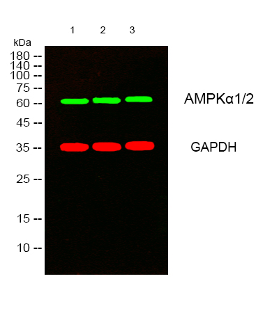
- Western blot analysis of lysates from 1) K562 , 2) COS7 , 3) KB cells, (Green) primary antibody was diluted at 1:1000, 4°over night, secondary antibody(cat:RS23920)was diluted at 1:10000, 37° 1hour. (Red) GAPDH Monoclonal Antibody(2B8) (cat:YM3029) antibody was diluted at 1:5000 as loading control, 4° over night,secondary antibody(cat:RS23710)was diluted at 1:10000, 37° 1hour.
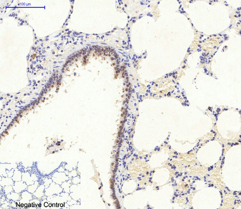
- Immunohistochemical analysis of paraffin-embedded Rat-lung tissue. 1,AMPKα1/2 Polyclonal Antibody was diluted at 1:200(4°C,overnight). 2, Sodium citrate pH 6.0 was used for antibody retrieval(>98°C,20min). 3,Secondary antibody was diluted at 1:200(room tempeRature, 30min). Negative control was used by secondary antibody only.
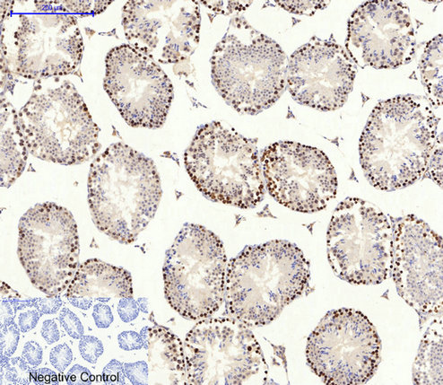
- Immunohistochemical analysis of paraffin-embedded Mouse-testis tissue. 1,AMPKα1/2 Polyclonal Antibody was diluted at 1:200(4°C,overnight). 2, Sodium citrate pH 6.0 was used for antibody retrieval(>98°C,20min). 3,Secondary antibody was diluted at 1:200(room tempeRature, 30min). Negative control was used by secondary antibody only.
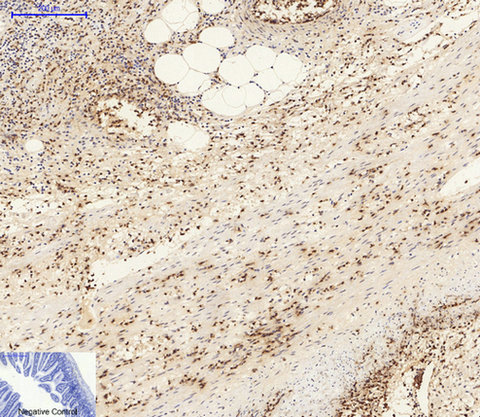
- Immunohistochemical analysis of paraffin-embedded Mouse-colon tissue. 1,AMPKα1/2 Polyclonal Antibody was diluted at 1:200(4°C,overnight). 2, Sodium citrate pH 6.0 was used for antibody retrieval(>98°C,20min). 3,Secondary antibody was diluted at 1:200(room tempeRature, 30min). Negative control was used by secondary antibody only.
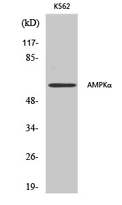
- Western Blot analysis of various cells using AMPKα1/2 Polyclonal Antibody diluted at 1:500
.jpg)
- Western Blot analysis of COS7 cells using AMPKα1/2 Polyclonal Antibody diluted at 1:500


