Akt1 Polyclonal Antibody
- Catalog No.:YT0177
- Applications:WB;IHC;IF;ELISA
- Reactivity:Human;Mouse;Rat
- Target:
- Akt1
- Fields:
- >>EGFR tyrosine kinase inhibitor resistance;>>Endocrine resistance;>>Platinum drug resistance;>>MAPK signaling pathway;>>ErbB signaling pathway;>>Ras signaling pathway;>>Rap1 signaling pathway;>>cGMP-PKG signaling pathway;>>cAMP signaling pathway;>>Chemokine signaling pathway;>>HIF-1 signaling pathway;>>FoxO signaling pathway;>>Sphingolipid signaling pathway;>>Phospholipase D signaling pathway;>>Autophagy - animal;>>mTOR signaling pathway;>>PI3K-Akt signaling pathway;>>AMPK signaling pathway;>>Apoptosis;>>Longevity regulating pathway;>>Longevity regulating pathway - multiple species;>>Cellular senescence;>>Adrenergic signaling in cardiomyocytes;>>VEGF signaling pathway;>>Apelin signaling pathway;>>Osteoclast differentiation;>>Focal adhesion;>>Signaling pathways regulating pluripotency of stem cells;>>Platelet activation;>>Neutrophil extracellular trap formation;>>Toll-like receptor signaling pathway;>>C-type lectin receptor signaling pathway;>>JAK-STAT signaling pathway;>>T cell recept
- Gene Name:
- AKT1
- Protein Name:
- RAC-alpha serine/threonine-protein kinase
- Human Gene Id:
- 207
- Human Swiss Prot No:
- P31749
- Mouse Gene Id:
- 11651
- Mouse Swiss Prot No:
- P31750
- Rat Gene Id:
- 24185
- Rat Swiss Prot No:
- P47196
- Immunogen:
- The antiserum was produced against synthesized peptide derived from human Akt. AA range:95-144
- Specificity:
- Akt1 Polyclonal Antibody detects endogenous levels of Akt1 protein.
- Formulation:
- Liquid in PBS containing 50% glycerol, 0.5% BSA and 0.02% sodium azide.
- Source:
- Polyclonal, Rabbit,IgG
- Dilution:
- WB 1:500 - 1:2000. IHC 1:100 - 1:300. IF 1:200 - 1:1000. ELISA: 1:40000. Not yet tested in other applications.
- Purification:
- The antibody was affinity-purified from rabbit antiserum by affinity-chromatography using epitope-specific immunogen.
- Concentration:
- 1 mg/ml
- Storage Stability:
- -15°C to -25°C/1 year(Do not lower than -25°C)
- Other Name:
- AKT1;PKB;RAC;RAC-alpha serine/threonine-protein kinase;Protein kinase B;PKB;Protein kinase B alpha;PKB alpha;Proto-oncogene c-Akt;RAC-PK-alpha
- Observed Band(KD):
- 56kD
- Background:
- The serine-threonine protein kinase encoded by the AKT1 gene is catalytically inactive in serum-starved primary and immortalized fibroblasts. AKT1 and the related AKT2 are activated by platelet-derived growth factor. The activation is rapid and specific, and it is abrogated by mutations in the pleckstrin homology domain of AKT1. It was shown that the activation occurs through phosphatidylinositol 3-kinase. In the developing nervous system AKT is a critical mediator of growth factor-induced neuronal survival. Survival factors can suppress apoptosis in a transcription-independent manner by activating the serine/threonine kinase AKT1, which then phosphorylates and inactivates components of the apoptotic machinery. Mutations in this gene have been associated with the Proteus syndrome. Multiple alternatively spliced transcript variants have been found for this gene. [provided by RefSeq, Jul 2011]
- Function:
- catalytic activity:ATP + a protein = ADP + a phosphoprotein.,disease:Defects in AKT1 are associated with breast cancer (BC) [MIM:114480]. BC is an extremely common malignancy, affecting one in eight women during their lifetime.,disease:Defects in AKT1 are associated with colorectal cancer (CRC) [MIM:114500].,disease:Defects in AKT1 are associated with susceptibility to ovarian cancer [MIM:604370]; also called susceptibility to familial breast-ovarian cancer type 1 (BROVCA1).,domain:Binding of the PH domain to the phosphatidylinositol 3-kinase alpha (PI(3)K) results in its targeting to the plasma membrane.,domain:The AGC-kinase C-terminal mediates interaction with THEM4.,enzyme regulation:Three specific sites, one in the kinase domain (Thr-308) and the two other ones in the C-terminal regulatory region (Ser-473 and Tyr-474), need to be phosphorylated for its full activation.,function:Gene
- Subcellular Location:
- Cytoplasm . Nucleus . Cell membrane . Nucleus after activation by integrin-linked protein kinase 1 (ILK1). Nuclear translocation is enhanced by interaction with TCL1A. Phosphorylation on Tyr-176 by TNK2 results in its localization to the cell membrane where it is targeted for further phosphorylations on Thr-308 and Ser-473 leading to its activation and the activated form translocates to the nucleus. Colocalizes with WDFY2 in intracellular vesicles (PubMed:16792529). .
- Expression:
- Expressed in prostate cancer and levels increase from the normal to the malignant state (at protein level). Expressed in all human cell types so far analyzed. The Tyr-176 phosphorylated form shows a significant increase in expression in breast cancers during the progressive stages i.e. normal to hyperplasia (ADH), ductal carcinoma in situ (DCIS), invasive ductal carcinoma (IDC) and lymph node metastatic (LNMM) stages.
Pregabalin Mediates Retinal Ganglion Cell Survival From Retinal Ischemia/Reperfusion Injury Via the Akt/GSK3β/β-Catenin Signaling Pathway
Insulin Attenuates Beta-Amyloid-Associated Insulin/Akt/EAAT Signaling Perturbations in Human Astrocytes. CELLULAR AND MOLECULAR NEUROBIOLOGY Cell Mol Neurobiol. 2016 Aug;36(6):851-864 WB Human 1:1000 Astrocytes
Over-Expression of Centromere Protein U Participates in the Malignant Neoplastic Progression of Breast Cancer. Frontiers in Oncology Front Oncol. 2021 Mar;0:350 WB Human 1:1000 Breast cancer tissues ,the paired adjacent histologically normal tissue T47D cell, MDA-MB-231 cell, SK-BR-3 cell, MCF-7 cell
Quantitative dot blot analysis (QDB), a versatile high throughput immunoblot method. Oncotarget Oncotarget. 2017 Aug 29; 8(35): 58553–58562 WB Human,Mouse liver HEK293 cell
Metallothionein-2 is associated with the amelioration of asthmatic pulmonary function by acupuncture through protein phosphorylation. BIOMEDICINE & PHARMACOTHERAPY 2019 Dec 23 WB Rat lung tissue
Zhang, Jiandi. "Apparatus and method for high trhoughput immunobloting." U.S. Patent Application No. 15/433,586.
- June 19-2018
- WESTERN IMMUNOBLOTTING PROTOCOL
- June 19-2018
- IMMUNOHISTOCHEMISTRY-PARAFFIN PROTOCOL
- June 19-2018
- IMMUNOFLUORESCENCE PROTOCOL
- September 08-2020
- FLOW-CYTOMEYRT-PROTOCOL
- May 20-2022
- Cell-Based ELISA│解您多样本WB检测之困扰
- July 13-2018
- CELL-BASED-ELISA-PROTOCOL-FOR-ACETYL-PROTEIN
- July 13-2018
- CELL-BASED-ELISA-PROTOCOL-FOR-PHOSPHO-PROTEIN
- July 13-2018
- Antibody-FAQs
- Products Images

- Immunofluorescence analysis of human-lung tissue. 1,Akt1 Polyclonal Antibody(red) was diluted at 1:200(4°C,overnight). 2, Cy3 labled Secondary antibody was diluted at 1:300(room temperature, 50min).3, Picture B: DAPI(blue) 10min. Picture A:Target. Picture B: DAPI. Picture C: merge of A+B

- Immunofluorescence analysis of human-lung tissue. 1,Akt1 Polyclonal Antibody(red) was diluted at 1:200(4°C,overnight). 2, Cy3 labled Secondary antibody was diluted at 1:300(room temperature, 50min).3, Picture B: DAPI(blue) 10min. Picture A:Target. Picture B: DAPI. Picture C: merge of A+B
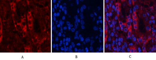
- Immunofluorescence analysis of human-stomach tissue. 1,Akt1 Polyclonal Antibody(red) was diluted at 1:200(4°C,overnight). 2, Cy3 labled Secondary antibody was diluted at 1:300(room temperature, 50min).3, Picture B: DAPI(blue) 10min. Picture A:Target. Picture B: DAPI. Picture C: merge of A+B

- Immunofluorescence analysis of human-stomach tissue. 1,Akt1 Polyclonal Antibody(red) was diluted at 1:200(4°C,overnight). 2, Cy3 labled Secondary antibody was diluted at 1:300(room temperature, 50min).3, Picture B: DAPI(blue) 10min. Picture A:Target. Picture B: DAPI. Picture C: merge of A+B
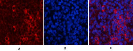
- Immunofluorescence analysis of rat-spleen tissue. 1,Akt1 Polyclonal Antibody(red) was diluted at 1:200(4°C,overnight). 2, Cy3 labled Secondary antibody was diluted at 1:300(room temperature, 50min).3, Picture B: DAPI(blue) 10min. Picture A:Target. Picture B: DAPI. Picture C: merge of A+B

- Immunofluorescence analysis of rat-spleen tissue. 1,Akt1 Polyclonal Antibody(red) was diluted at 1:200(4°C,overnight). 2, Cy3 labled Secondary antibody was diluted at 1:300(room temperature, 50min).3, Picture B: DAPI(blue) 10min. Picture A:Target. Picture B: DAPI. Picture C: merge of A+B
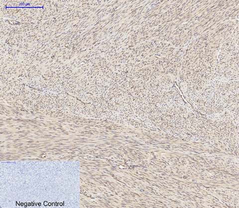
- Immunohistochemical analysis of paraffin-embedded Human-uterus tissue. 1,Akt1 Polyclonal Antibody was diluted at 1:200(4°C,overnight). 2, Sodium citrate pH 6.0 was used for antibody retrieval(>98°C,20min). 3,Secondary antibody was diluted at 1:200(room tempeRature, 30min). Negative control was used by secondary antibody only.

- Immunohistochemical analysis of paraffin-embedded Human-uterus-cancer tissue. 1,Akt1 Polyclonal Antibody was diluted at 1:200(4°C,overnight). 2, Sodium citrate pH 6.0 was used for antibody retrieval(>98°C,20min). 3,Secondary antibody was diluted at 1:200(room tempeRature, 30min). Negative control was used by secondary antibody only.
- Western Blot analysis of various cells using Akt1 Polyclonal Antibody diluted at 1:2000
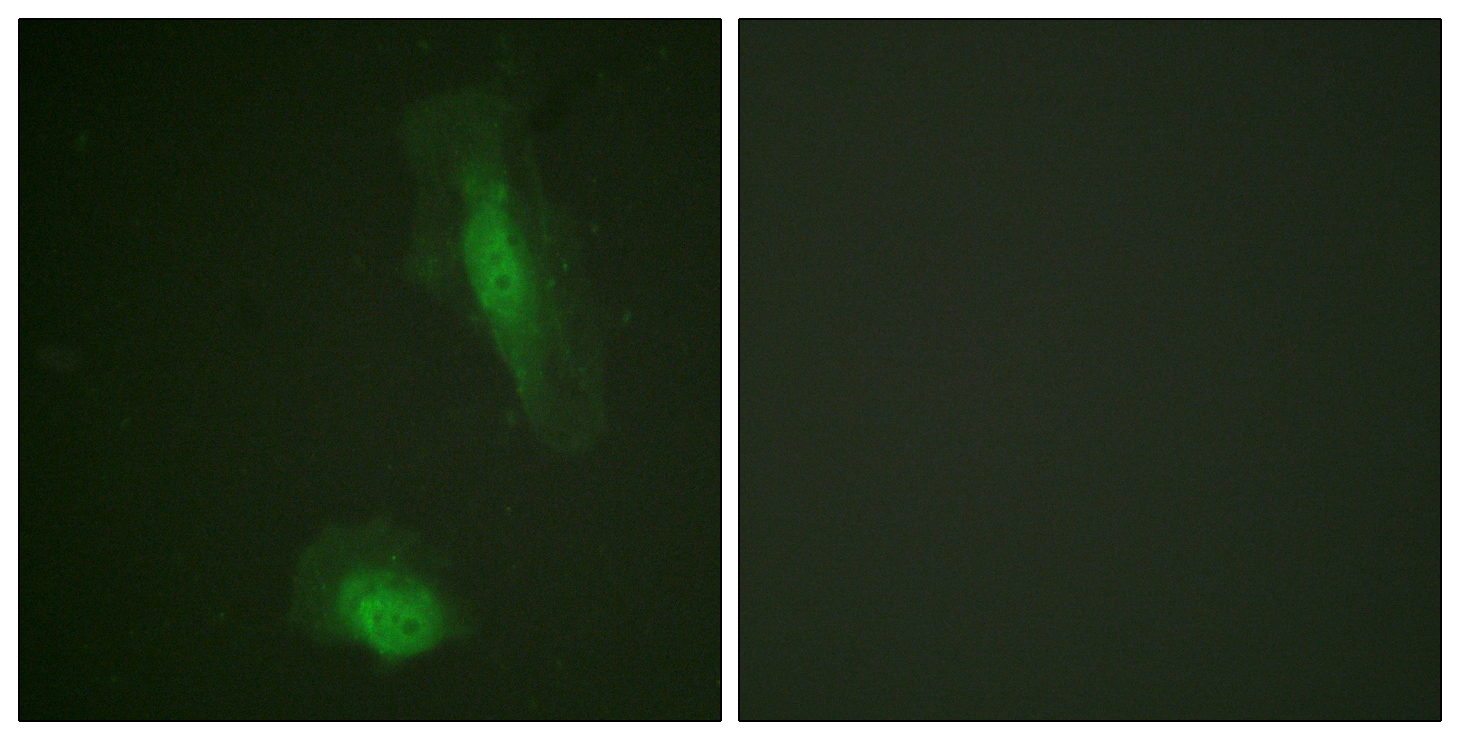
- Immunofluorescence analysis of HeLa cells, using Akt Antibody. The picture on the right is blocked with the synthesized peptide.
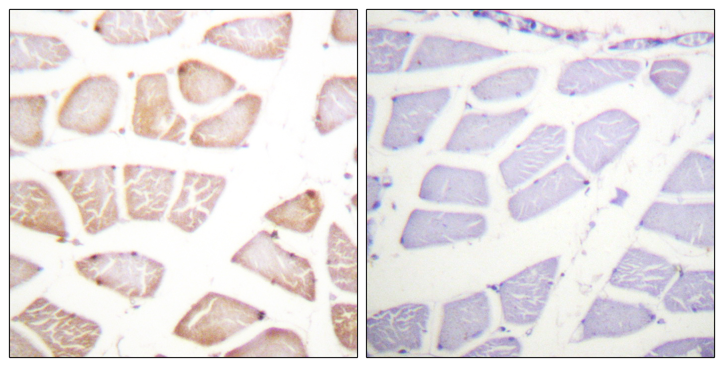
- Immunohistochemistry analysis of paraffin-embedded human skeletal muscle tissue, using Akt Antibody. The picture on the right is blocked with the synthesized peptide.
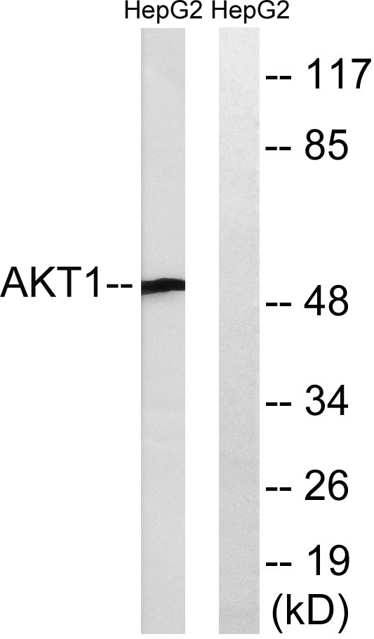
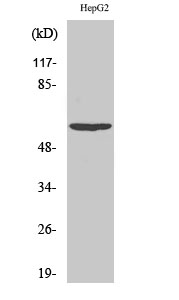
- Western blot analysis of lysates from HepG2 cells, treated with Serum 30% 30', using Akt Antibody. The lane on the right is blocked with the synthesized peptide.


