Syk (phospho Tyr323) Polyclonal Antibody
- Catalog No.:YP0896
- Applications:WB;IHC;IF;ELISA
- Reactivity:Human;Mouse;Rat
- Target:
- Syk
- Fields:
- >>NF-kappa B signaling pathway;>>Phospholipase D signaling pathway;>>PI3K-Akt signaling pathway;>>Osteoclast differentiation;>>Platelet activation;>>Neutrophil extracellular trap formation;>>C-type lectin receptor signaling pathway;>>Natural killer cell mediated cytotoxicity;>>B cell receptor signaling pathway;>>Fc epsilon RI signaling pathway;>>Fc gamma R-mediated phagocytosis;>>Tuberculosis;>>Kaposi sarcoma-associated herpesvirus infection;>>Herpes simplex virus 1 infection;>>Epstein-Barr virus infection;>>Coronavirus disease - COVID-19;>>Viral carcinogenesis
- Gene Name:
- SYK
- Protein Name:
- Tyrosine-protein kinase SYK
- Human Gene Id:
- 6850
- Human Swiss Prot No:
- P43405
- Mouse Gene Id:
- 20963
- Mouse Swiss Prot No:
- P48025
- Rat Gene Id:
- 25155
- Rat Swiss Prot No:
- Q64725
- Immunogen:
- The antiserum was produced against synthesized peptide derived from human SYK around the phosphorylation site of Tyr323. AA range:289-338
- Specificity:
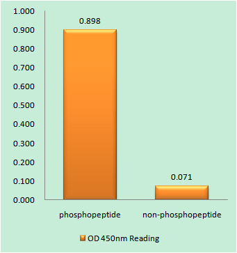
- Phospho-Syk (Y323) Polyclonal Antibody detects endogenous levels of Syk protein only when phosphorylated at Y323.
- Formulation:
- Liquid in PBS containing 50% glycerol, 0.5% BSA and 0.02% sodium azide.
- Source:
- Polyclonal, Rabbit,IgG
- Dilution:
- WB 1:500 - 1:2000. IHC 1:100 - 1:300. IF 1:200 - 1:1000. ELISA: 1:5000. Not yet tested in other applications.
- Purification:
- The antibody was affinity-purified from rabbit antiserum by affinity-chromatography using epitope-specific immunogen.
- Concentration:
- 1 mg/ml
- Storage Stability:
- -15°C to -25°C/1 year(Do not lower than -25°C)
- Other Name:
- SYK;Tyrosine-protein kinase SYK;Spleen tyrosine kinase;p72-Syk
- Observed Band(KD):
- 80kD
- Background:
- This gene encodes a member of the family of non-receptor type Tyr protein kinases. This protein is widely expressed in hematopoietic cells and is involved in coupling activated immunoreceptors to downstream signaling events that mediate diverse cellular responses, including proliferation, differentiation, and phagocytosis. It is thought to be a modulator of epithelial cell growth and a potential tumour suppressor in human breast carcinomas. Alternatively spliced transcript variants encoding different isoforms have been found for this gene. [provided by RefSeq, Mar 2010],
- Function:
- catalytic activity:ATP + a [protein]-L-tyrosine = ADP + a [protein]-L-tyrosine phosphate.,function:Positive effector of BCR-stimulated responses. Couples the B-cell antigen receptor (BCR) to the mobilization of calcium ion either through a phosphoinositide 3-kinase-dependent pathway, when not phosphorylated on tyrosines of the linker region, or through a phospholipase C-gamma-dependent pathway, when phosphorylated on Tyr-348 and Tyr-352. Thus the differential phosphorylation of Syk can determine the pathway by which BCR is coupled to the regulation of intracellular calcium ion.,PTM:Autophosphorylated.,PTM:Phosphorylation on Tyr-323 creates a binding site for c-Cbl, an adapter protein that serves as a negative regulator of BCR-stimulated calcium ion signaling.,PTM:Phosphorylation on Tyr-348 and Tyr-352 enhances the phosphorylation and activation of phospholipase C-gamma and the early phas
- Subcellular Location:
- Cell membrane . Cytoplasm, cytosol .
- Expression:
- Widely expressed in hematopoietic cells (at protein level) (PubMed:8163536). Expressed in neutrophils (at protein level) (PubMed:15123770). Within the B-cell compartment, expressed from pro- and pre-B cells to plasma cells (PubMed:8163536).
- June 19-2018
- WESTERN IMMUNOBLOTTING PROTOCOL
- June 19-2018
- IMMUNOHISTOCHEMISTRY-PARAFFIN PROTOCOL
- June 19-2018
- IMMUNOFLUORESCENCE PROTOCOL
- September 08-2020
- FLOW-CYTOMEYRT-PROTOCOL
- May 20-2022
- Cell-Based ELISA│解您多样本WB检测之困扰
- July 13-2018
- CELL-BASED-ELISA-PROTOCOL-FOR-ACETYL-PROTEIN
- July 13-2018
- CELL-BASED-ELISA-PROTOCOL-FOR-PHOSPHO-PROTEIN
- July 13-2018
- Antibody-FAQs
- Products Images

- Immunofluorescence analysis of HepG2 cells, using SYK (Phospho-Tyr323) Antibody. The picture on the right is blocked with the phospho peptide.

- Immunohistochemistry analysis of paraffin-embedded human lymph node, using SYK (Phospho-Tyr323) Antibody. The picture on the right is blocked with the phospho peptide.

- Western blot analysis of lysates from HT29 cells, using SYK (Phospho-Tyr323) Antibody. The lane on the right is blocked with the phospho peptide.