PI 3 kinase p85/p55 (phospho Tyr467/199) Polyclonal Antibody
- Catalog No.:YP0224
- Applications:WB;IHC;IF;ELISA
- Reactivity:Human;Mouse;Rat;Monkey;Pig
- Target:
- PI3 kinase p85/p55
- Fields:
- >>EGFR tyrosine kinase inhibitor resistance;>>Endocrine resistance;>>Platinum drug resistance;>>ErbB signaling pathway;>>Ras signaling pathway;>>Rap1 signaling pathway;>>cAMP signaling pathway;>>Chemokine signaling pathway;>>HIF-1 signaling pathway;>>FoxO signaling pathway;>>Phosphatidylinositol signaling system;>>Sphingolipid signaling pathway;>>Phospholipase D signaling pathway;>>Autophagy - animal;>>mTOR signaling pathway;>>PI3K-Akt signaling pathway;>>AMPK signaling pathway;>>Apoptosis;>>Longevity regulating pathway;>>Longevity regulating pathway - multiple species;>>Cellular senescence;>>Axon guidance;>>VEGF signaling pathway;>>Osteoclast differentiation;>>Focal adhesion;>>Signaling pathways regulating pluripotency of stem cells;>>Platelet activation;>>Neutrophil extracellular trap formation;>>Toll-like receptor signaling pathway;>>C-type lectin receptor signaling pathway;>>JAK-STAT signaling pathway;>>Natural killer cell mediated cytotoxicity;>>T cell receptor signaling pathway;>
- Gene Name:
- PIK3R1/PIK3R3
- Protein Name:
- Phosphatidylinositol 3-kinase regulatory subunit alpha/gamma
- Human Gene Id:
- 5295
- Human Swiss Prot No:
- P27986/Q92569
- Mouse Gene Id:
- 18708/18710
- Rat Gene Id:
- 25513/60664
- Rat Swiss Prot No:
- Q63787/Q63789
- Immunogen:
- The antiserum was produced against synthesized peptide derived from human PI3-kinase p85-alpha/gamma around the phosphorylation site of Tyr467/199. AA range:436-485
- Specificity:
- Phospho-PI 3-kinase p85/p55 (Y467/199) Polyclonal Antibody detects endogenous levels of PI 3-kinase p85/p55 protein only when phosphorylated at Y467/199.
- Formulation:
- Liquid in PBS containing 50% glycerol, 0.5% BSA and 0.02% sodium azide.
- Source:
- Polyclonal, Rabbit,IgG
- Dilution:
- WB 1:500 - 1:2000. IHC 1:100 - 1:300. IF 1:200 - 1:1000. ELISA: 1:10000. Not yet tested in other applications.
- Purification:
- The antibody was affinity-purified from rabbit antiserum by affinity-chromatography using epitope-specific immunogen.
- Concentration:
- 1 mg/ml
- Storage Stability:
- -15°C to -25°C/1 year(Do not lower than -25°C)
- Other Name:
- PIK3R1;GRB1;Phosphatidylinositol 3-kinase regulatory subunit alpha;PI3-kinase regulatory subunit alpha;PI3K regulatory subunit alpha;PtdIns-3-kinase regulatory subunit alpha;Phosphatidylinositol 3-kinase 85 kDa regulatory subunit alpha;PI3-kinase subunit p85-alpha;PtdIns-3-kinase regulatory subunit p85-alpha
- Observed Band(KD):
- 55+85kD
- Background:
- Phosphatidylinositol 3-kinase phosphorylates the inositol ring of phosphatidylinositol at the 3-prime position. The enzyme comprises a 110 kD catalytic subunit and a regulatory subunit of either 85, 55, or 50 kD. This gene encodes the 85 kD regulatory subunit. Phosphatidylinositol 3-kinase plays an important role in the metabolic actions of insulin, and a mutation in this gene has been associated with insulin resistance. Alternative splicing of this gene results in four transcript variants encoding different isoforms. [provided by RefSeq, Jun 2011],
- Function:
- disease:Defects in PIK3R1 are a cause of severe insulin resistance.,domain:The SH3 domain mediates the binding to CBLB, and to HIV-1 Nef.,function:Binds to activated (phosphorylated) protein-Tyr kinases, through its SH2 domain, and acts as an adapter, mediating the association of the p110 catalytic unit to the plasma membrane. Necessary for the insulin-stimulated increase in glucose uptake and glycogen synthesis in insulin-sensitive tissues.,PTM:Polyubiquitinated in T-cells by CBLB; which does not promote proteasomal degradation but impairs association with CD28 and CD3Z upon T-cell activation.,similarity:Belongs to the PI3K p85 subunit family.,similarity:Contains 1 Rho-GAP domain.,similarity:Contains 1 SH3 domain.,similarity:Contains 2 SH2 domains.,subunit:Heterodimer of a p110 (catalytic) and a p85 (regulatory) subunits. Interacts with phosphorylated TOM1L1. Interacts with phosphorylat
- Subcellular Location:
- nucleus,cytoplasm,cis-Golgi network,cytosol,plasma membrane,cell-cell junction,phosphatidylinositol 3-kinase complex,phosphatidylinositol 3-kinase complex, class IA,membrane,perinuclear endoplasmic reticulum membrane,
- Expression:
- Isoform 2 is expressed in skeletal muscle and brain, and at lower levels in kidney and cardiac muscle. Isoform 2 and isoform 4 are present in skeletal muscle (at protein level).
Maltol ameliorates intervertebral disc degeneration through inhibiting PI3K/AKT/NF-κB pathway and regulating NLRP3 inflammasome-mediated pyroptosis
Network Pharmacology Analysis and Experimental Validation to Investigate the Mechanism of Total Flavonoids of Rhizoma Drynariae in Treating Rheumatoid Arthritis WB Rat /RA-FLSs
Integrating Network Pharmacology and Experimental Validation to Explore the Key Mechanism of Gubitong Recipe in the Treatment of Osteoarthritis WB Human 1 : 1000 /SW1353 cells
An Integrated Approach Based on Network Analysis Combined With Experimental Verification Reveals PI3K/Akt/Nrf2 Signaling Is an Important Way for the Anti-Myocardial Ischemia Activity of Yi-Qi-Tong-Luo Capsule. Front Pharmacol. 2022 Feb;13:794528-794528. WB,IF Rat 1:1000
TSSK4 upregulation in alveolar epithelial type-II cells facilitates pulmonary fibrosis through HSP90-AKT signaling restriction and AT-II apoptosis. Cell Death & Disease Cell Death Dis. 2021 Oct;12(10):1-14 WB Mouse MLE12 cell
Regulatory effects of hawthorn polyphenols on hyperglycemic, inflammatory, insulin resistance responses, and alleviation of aortic injury in type 2 diabetic rats. FOOD RESEARCH INTERNATIONAL Food Res Int. 2021 Apr;142:110239 WB Rat 1:1000 Quadriceps,liver,aorta
Rapamycin Antagonizes BCRP-Mediated Drug Resistance Through the PI3K/Akt/mTOR Signaling Pathway in mPRα-Positive Breast Cancer. Frontiers in Oncology Front Oncol. 2021 Apr;0:352 WB Human 1 : 500 MDA-MB-231/BCRP cell,MDA-MB-453/BCRP cell
5‐FU inhibits migration and invasion of CRC cells through PI3K/AKT pathway regulated by MARCH1. CELL BIOLOGY INTERNATIONAL Cell Biol Int. 2021 Feb;45(2):368-381 WB Human Colorectal cancer(CRC ) tissues sw480s,DLD-1s,NCM460s
Polygonum cuspidatum Extract Exerts Antihyperlipidemic Effects by Regulation of PI3K/AKT/FOXO3 Signaling Pathway. Oxidative Medicine and Cellular Longevity Oxid Med Cell Longev. 2021;2021:3830671 WB Human 1:1000 HepG2 cells
Hydroxy-α-sanshool Possesses Protective Potentials on H2O2-Stimulated PC12 Cells by Suppression of Oxidative Stress-Induced Apoptosis through Regulation of PI3K/Akt Signal Pathway. Oxidative Medicine and Cellular Longevity Oxid Med Cell Longev. 2020;2020:3481758 WB Rat 1 : 1000 PC12 cell
Lapatinib‑induced inhibition of ovarian function is counteracted by the STAT3 pathway both in vivo and in vitro. ONCOLOGY REPORTS Oncol Rep. 2020 Sep;44(3):1127-1135 IHC Mouse 1:50 Ovarian tissue Oocytes
Deficiency in the short‐chain acyl‐CoA dehydrogenase protects mice against diet‐induced obesity and insulin resistance. FASEB JOURNAL Faseb J. 2019 Dec;33(12):13722-13733 WB Mouse liver hepatocytes
The role and regulatory mechanism of autophagy in hippocampal nerve cells of piglet damaged by deoxynivalenol. TOXICOLOGY IN VITRO Toxicol In Vitro. 2020 Aug;66:104837 WB Pig 1:1000 piglet hippocampal nerve cells (PHNCs)
Wang, Wei-wei, et al. "Telmisartan reduces atrial arrhythmia susceptibility through the regulation of RAS–ERK and PI3K–Akt–eNOS pathways in spontaneously hypertensive rats." Canadian journal of physiology and pharmacology 93.8 (2015): 657-665.
佟雪, et al. "基于 PI3K/AKT/mTOR 信号通路研究补肾法对体外培养小鼠卵母细胞成熟的促进作用." 北京中医药大学学报 44.6 (2021): 510-518.
恪雪, et al. "基于 PI3K/AKT/mTOR 信号通路研究补肾法对体外培养 小鼠卵母细胞成熟的促进作." Journal of Beijing University of Traditional Chinese Medicine 44.6 (2021).
Chondroprotective Effects of Gubitong Recipe via Inhibiting Excessive Mitophagy of Chondrocytes Evidence-based Complementary and Alternative Medicine Jing Luo WB Rat
The adhesion protein of Mycoplasma genitalium inhibits urethral epithelial cell apoptosis through CypA-CD147 activating PI3K/ Akt/NF-κB pathway APPLIED MICROBIOLOGY AND BIOTECHNOLOGY Yanhua Zeng WB Human
Network and Experimental Pharmacology to Decode the Action of Wendan Decoction Against Generalized Anxiety Disorder Drug Design Development and Therapy Xin Zhao WB Mouse
Effect of STK3 on proliferation and apoptosis of pancreatic cancer cells via PI3K/AKT/mTOR pathway CELLULAR SIGNALLING Zheng Jiang WB Mouse,Human PANC-1 cell-xenograft,empty vector cell-xenograft BXPC-3 cell, PANC-1cell
AMPK/mTOR/p70S6K axis prevents apoptosis of Porphyromonas gingivalis-infected gingival epithelial cells via BadSer136 phosphorylation APOPTOSIS He Hongbing WB Human human gingival epithelial cells (hGECs)
Carbon Ions Suppress Angiogenesis and Lung Metastases in Melanoma by Targeting CXCL10. RADIATION RESEARCH Chengcheng Li WB Mouse 1: 1000 melanoma tissues
Quercetin improves cerebral ischemia/reperfusion injury by promoting microglia/macrophages M2 polarization via regulating PI3K/Akt/NF-κB signaling pathway. BIOMEDICINE & PHARMACOTHERAPY Lisheng Chu WB Rat Microglia
Cucurbitacin E reduces IL-1β-induced inflammation and cartilage degeneration by inhibiting the PI3K/Akt pathway in osteoarthritic chondrocytes Journal of Translational Medicine Hui Zhang WB Human 1:1000 Chondrocytes
- June 19-2018
- WESTERN IMMUNOBLOTTING PROTOCOL
- June 19-2018
- IMMUNOHISTOCHEMISTRY-PARAFFIN PROTOCOL
- June 19-2018
- IMMUNOFLUORESCENCE PROTOCOL
- September 08-2020
- FLOW-CYTOMEYRT-PROTOCOL
- May 20-2022
- Cell-Based ELISA│解您多样本WB检测之困扰
- July 13-2018
- CELL-BASED-ELISA-PROTOCOL-FOR-ACETYL-PROTEIN
- July 13-2018
- CELL-BASED-ELISA-PROTOCOL-FOR-PHOSPHO-PROTEIN
- July 13-2018
- Antibody-FAQs
- Products Images
- Yang, Yazong, et al. "Fucoidan inhibits lymphangiogenesis by downregulating the expression of VEGFR3 and PROX1 in human lymphatic endothelial cells." Oncotarget 7.25 (2016): 38025.
-if-94.jpg)
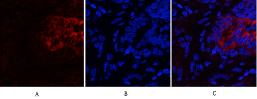
- Immunofluorescence analysis of human-lung tissue. 1,PI 3-kinase p85/p55 (phospho Tyr467/199) Polyclonal Antibody(red) was diluted at 1:200(4°C,overnight). 2, Cy3 labled Secondary antibody was diluted at 1:300(room temperature, 50min).3, Picture B: DAPI(blue) 10min. Picture A:Target. Picture B: DAPI. Picture C: merge of A+B
-if-95.jpg)
- Immunofluorescence analysis of human-lung tissue. 1,PI 3-kinase p85/p55 (phospho Tyr467/199) Polyclonal Antibody(red) was diluted at 1:200(4°C,overnight). 2, Cy3 labled Secondary antibody was diluted at 1:300(room temperature, 50min).3, Picture B: DAPI(blue) 10min. Picture A:Target. Picture B: DAPI. Picture C: merge of A+B
-if-96.jpg)
- Immunofluorescence analysis of human-stomach tissue. 1,PI 3-kinase p85/p55 (phospho Tyr467/199) Polyclonal Antibody(red) was diluted at 1:200(4°C,overnight). 2, Cy3 labled Secondary antibody was diluted at 1:300(room temperature, 50min).3, Picture B: DAPI(blue) 10min. Picture A:Target. Picture B: DAPI. Picture C: merge of A+B
-if-97.jpg)
- Immunofluorescence analysis of human-stomach tissue. 1,PI 3-kinase p85/p55 (phospho Tyr467/199) Polyclonal Antibody(red) was diluted at 1:200(4°C,overnight). 2, Cy3 labled Secondary antibody was diluted at 1:300(room temperature, 50min).3, Picture B: DAPI(blue) 10min. Picture A:Target. Picture B: DAPI. Picture C: merge of A+B
-if-98.jpg)
- Immunofluorescence analysis of rat-spleen tissue. 1,PI 3-kinase p85/p55 (phospho Tyr467/199) Polyclonal Antibody(red) was diluted at 1:200(4°C,overnight). 2, Cy3 labled Secondary antibody was diluted at 1:300(room temperature, 50min).3, Picture B: DAPI(blue) 10min. Picture A:Target. Picture B: DAPI. Picture C: merge of A+B
-if-99.jpg)
- Immunofluorescence analysis of rat-spleen tissue. 1,PI 3-kinase p85/p55 (phospho Tyr467/199) Polyclonal Antibody(red) was diluted at 1:200(4°C,overnight). 2, Cy3 labled Secondary antibody was diluted at 1:300(room temperature, 50min).3, Picture B: DAPI(blue) 10min. Picture A:Target. Picture B: DAPI. Picture C: merge of A+B
2016-11-25.jpg)
- Western Blot analysis of various cells using primary antibody diluted at 1:1000(4°C overnight). Secondary antibody:Goat Anti-rabbit IgG IRDye 800( diluted at 1:5000, 25°C, 1 hour)
poly-ihc-human-uterus.jpg)
- Immunohistochemical analysis of paraffin-embedded Human-uterus tissue. 1,PI 3-kinase p85/p55 (phospho Tyr467/199) Polyclonal Antibody was diluted at 1:200(4°C,overnight). 2, Sodium citrate pH 6.0 was used for antibody retrieval(>98°C,20min). 3,Secondary antibody was diluted at 1:200(room tempeRature, 30min). Negative control was used by secondary antibody only.
poly-ihc-human-colon.jpg)
- Immunohistochemical analysis of paraffin-embedded Human-colon tissue. 1,PI 3-kinase p85/p55 (phospho Tyr467/199) Polyclonal Antibody was diluted at 1:200(4°C,overnight). 2, Sodium citrate pH 6.0 was used for antibody retrieval(>98°C,20min). 3,Secondary antibody was diluted at 1:200(room tempeRature, 30min). Negative control was used by secondary antibody only.
poly-ihc-human-liver.jpg)
- Immunohistochemical analysis of paraffin-embedded Human-liver tissue. 1,PI 3-kinase p85/p55 (phospho Tyr467/199) Polyclonal Antibody was diluted at 1:200(4°C,overnight). 2, Sodium citrate pH 6.0 was used for antibody retrieval(>98°C,20min). 3,Secondary antibody was diluted at 1:200(room tempeRature, 30min). Negative control was used by secondary antibody only.
poly-ihc-human-lung.jpg)
- Immunohistochemical analysis of paraffin-embedded Human-lung tissue. 1,PI 3-kinase p85/p55 (phospho Tyr467/199) Polyclonal Antibody was diluted at 1:200(4°C,overnight). 2, Sodium citrate pH 6.0 was used for antibody retrieval(>98°C,20min). 3,Secondary antibody was diluted at 1:200(room tempeRature, 30min). Negative control was used by secondary antibody only.
poly-ihc-human-stomach-cancer.jpg)
- Immunohistochemical analysis of paraffin-embedded Human-stomach-cancer tissue. 1,PI 3-kinase p85/p55 (phospho Tyr467/199) Polyclonal Antibody was diluted at 1:200(4°C,overnight). 2, Sodium citrate pH 6.0 was used for antibody retrieval(>98°C,20min). 3,Secondary antibody was diluted at 1:200(room tempeRature, 30min). Negative control was used by secondary antibody only.
poly-ihc-rat-heart.jpg)
- Immunohistochemical analysis of paraffin-embedded Rat-heart tissue. 1,PI 3-kinase p85/p55 (phospho Tyr467/199) Polyclonal Antibody was diluted at 1:200(4°C,overnight). 2, Sodium citrate pH 6.0 was used for antibody retrieval(>98°C,20min). 3,Secondary antibody was diluted at 1:200(room tempeRature, 30min). Negative control was used by secondary antibody only.
poly-ihc-rat-liver.jpg)
- Immunohistochemical analysis of paraffin-embedded Rat-liver tissue. 1,PI 3-kinase p85/p55 (phospho Tyr467/199) Polyclonal Antibody was diluted at 1:200(4°C,overnight). 2, Sodium citrate pH 6.0 was used for antibody retrieval(>98°C,20min). 3,Secondary antibody was diluted at 1:200(room tempeRature, 30min). Negative control was used by secondary antibody only.
poly-ihc-rat-lung.jpg)
- Immunohistochemical analysis of paraffin-embedded Rat-lung tissue. 1,PI 3-kinase p85/p55 (phospho Tyr467/199) Polyclonal Antibody was diluted at 1:200(4°C,overnight). 2, Sodium citrate pH 6.0 was used for antibody retrieval(>98°C,20min). 3,Secondary antibody was diluted at 1:200(room tempeRature, 30min). Negative control was used by secondary antibody only.
poly-ihc-rat-kidney.jpg)
- Immunohistochemical analysis of paraffin-embedded Rat-kidney tissue. 1,PI 3-kinase p85/p55 (phospho Tyr467/199) Polyclonal Antibody was diluted at 1:200(4°C,overnight). 2, Sodium citrate pH 6.0 was used for antibody retrieval(>98°C,20min). 3,Secondary antibody was diluted at 1:200(room tempeRature, 30min). Negative control was used by secondary antibody only.
poly-ihc-rat-spinal-cord.jpg)
- Immunohistochemical analysis of paraffin-embedded Rat-spinal-cord tissue. 1,PI 3-kinase p85/p55 (phospho Tyr467/199) Polyclonal Antibody was diluted at 1:200(4°C,overnight). 2, Sodium citrate pH 6.0 was used for antibody retrieval(>98°C,20min). 3,Secondary antibody was diluted at 1:200(room tempeRature, 30min). Negative control was used by secondary antibody only.
poly-ihc-rat-brain.jpg)
- Immunohistochemical analysis of paraffin-embedded Rat-brain tissue. 1,PI 3-kinase p85/p55 (phospho Tyr467/199) Polyclonal Antibody was diluted at 1:200(4°C,overnight). 2, Sodium citrate pH 6.0 was used for antibody retrieval(>98°C,20min). 3,Secondary antibody was diluted at 1:200(room tempeRature, 30min). Negative control was used by secondary antibody only.
poly-ihc-mouse-heart.jpg)
- Immunohistochemical analysis of paraffin-embedded Mouse-heart tissue. 1,PI 3-kinase p85/p55 (phospho Tyr467/199) Polyclonal Antibody was diluted at 1:200(4°C,overnight). 2, Sodium citrate pH 6.0 was used for antibody retrieval(>98°C,20min). 3,Secondary antibody was diluted at 1:200(room tempeRature, 30min). Negative control was used by secondary antibody only.
poly-ihc-mouse-testis.jpg)
- Immunohistochemical analysis of paraffin-embedded Mouse-testis tissue. 1,PI 3-kinase p85/p55 (phospho Tyr467/199) Polyclonal Antibody was diluted at 1:200(4°C,overnight). 2, Sodium citrate pH 6.0 was used for antibody retrieval(>98°C,20min). 3,Secondary antibody was diluted at 1:200(room tempeRature, 30min). Negative control was used by secondary antibody only.
poly-ihc-mouse-kidney.jpg)
- Immunohistochemical analysis of paraffin-embedded Mouse-kidney tissue. 1,PI 3-kinase p85/p55 (phospho Tyr467/199) Polyclonal Antibody was diluted at 1:200(4°C,overnight). 2, Sodium citrate pH 6.0 was used for antibody retrieval(>98°C,20min). 3,Secondary antibody was diluted at 1:200(room tempeRature, 30min). Negative control was used by secondary antibody only.
poly-ihc-mouse-brain.jpg)
- Immunohistochemical analysis of paraffin-embedded Mouse-brain tissue. 1,PI 3-kinase p85/p55 (phospho Tyr467/199) Polyclonal Antibody was diluted at 1:200(4°C,overnight). 2, Sodium citrate pH 6.0 was used for antibody retrieval(>98°C,20min). 3,Secondary antibody was diluted at 1:200(room tempeRature, 30min). Negative control was used by secondary antibody only.
- Western Blot analysis of COS7 cells using Phospho-PI 3-kinase p85/p55 (Y467/199) Polyclonal Antibody diluted at 1:1000
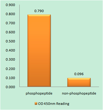
- Enzyme-Linked Immunosorbent Assay (Phospho-ELISA) for Immunogen Phosphopeptide (Phospho-left) and Non-Phosphopeptide (Phospho-right), using PI3-kinase p85-alpha/gamma (Phospho-Tyr467/199) Antibody
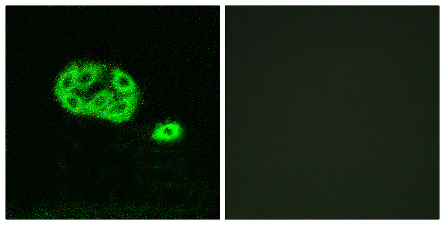
- Immunofluorescence analysis of NIH/3T3 cells, using PI3-kinase p85-alpha/gamma (Phospho-Tyr467/199) Antibody. The picture on the right is blocked with the phospho peptide.
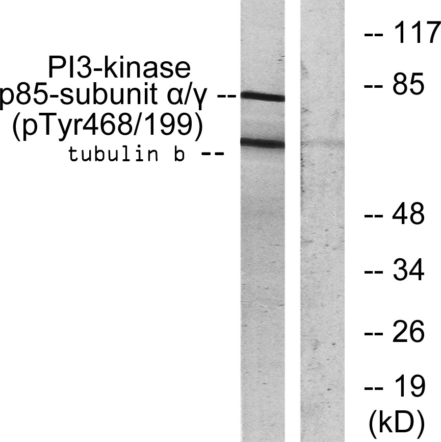
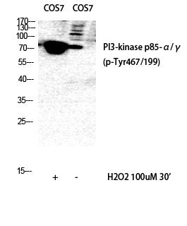
- Western blot analysis of lysates from COS7 cells treated with H2O2 100uM 30', using PI3-kinase p85-alpha/gamma (Phospho-Tyr467/199) Antibody. The lane on the right is blocked with the phospho peptide.