BEST2 Polyclonal Antibody
- Catalog No.:YN5663
- Applications:WB;IHC;IF
- Reactivity:Mouse;Rat
- Target:
- BEST2
- Fields:
- >>Salivary secretion
- Gene Name:
- BEST2
- Protein Name:
- Bestrophin-2
- Human Gene Id:
- 54831
- Human Swiss Prot No:
- Q8NFU1
- Mouse Swiss Prot No:
- Q8BGM5
- Immunogen:
- Synthetic Peptide of BEST2
- Specificity:
- The antibody detects endogenous BEST2 protein.
- Formulation:
- PBS, pH 7.4, containing 0.5%BSA, 0.02% sodium azide as Preservative and 50% Glycerol.
- Source:
- Polyclonal, Rabbit,IgG
- Dilution:
- WB 1:500-1000 IHC 1:200-500. IF 1:50-200
- Purification:
- The antibody was affinity-purified from rabbit antiserum by affinity-chromatography using epitope-specific immunogen.
- Storage Stability:
- -15°C to -25°C/1 year(Do not lower than -25°C)
- Other Name:
- Bestrophin-2;Vitelliform macular dystrophy 2-like protein 1
- Observed Band(KD):
- 50kD
- Background:
- This gene is a member of the bestrophin gene family of anion channels. Bestrophin genes share a similar gene structure with highly conserved exon-intron boundaries, but with distinct 3' ends. Bestrophins are transmembrane proteins that contain a homologous region rich in aromatic residues, including an invariant arg-phe-pro motif. Mutation in one of the family members (bestrophin 1) is associated with vitelliform macular dystrophy. The bestrophin 2 gene is mainly expressed in the retinal pigment epithelium and colon. [provided by RefSeq, Jul 2008],
- Function:
- function:Forms calcium-sensitive chloride channels. May conduct other physiologically significant anions such as bicarbonate.,similarity:Belongs to the bestrophin family.,tissue specificity:Mainly confined to the retinal pigment epithelium and colon.,
- Subcellular Location:
- Cell membrane; Multi-pass membrane protein.
- Expression:
- Mainly confined to the retinal pigment epithelium and colon.
- June 19-2018
- WESTERN IMMUNOBLOTTING PROTOCOL
- June 19-2018
- IMMUNOHISTOCHEMISTRY-PARAFFIN PROTOCOL
- June 19-2018
- IMMUNOFLUORESCENCE PROTOCOL
- September 08-2020
- FLOW-CYTOMEYRT-PROTOCOL
- May 20-2022
- Cell-Based ELISA│解您多样本WB检测之困扰
- July 13-2018
- CELL-BASED-ELISA-PROTOCOL-FOR-ACETYL-PROTEIN
- July 13-2018
- CELL-BASED-ELISA-PROTOCOL-FOR-PHOSPHO-PROTEIN
- July 13-2018
- Antibody-FAQs
- Products Images
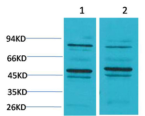
- Western blot analysis of 1) Mouse Brain Tissue, 2) Rat Brain Tissue using BEST2 Polyclonal Antibody. Secondary antibody(catalog#:RS0002) was diluted at 1:20000
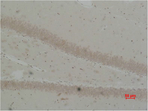
- Immunohistochemical analysis of paraffin-embedded Rat Brain Tissue using BEST2 Polyclonal Antibody.


