Melan-A (ABT-Mart1) Mouse mAb
- Catalog No.:YM6702
- Applications:WB;IHC;ELISA
- Reactivity:Human (predicted: Mouse)
- Target:
- Melan-A
- Gene Name:
- MLANA MART1
- Protein Name:
- Mart-1/Melan-A
- Human Gene Id:
- 2315
- Human Swiss Prot No:
- Q16655
- Immunogen:
- Synthesized peptide derived from human Mart-1/Melan-A AA range: 50-118
- Specificity:
- This antibody detects endogenous levels of human Mart-1/Melan-A. Heat-induced epitope retrieval (HIER) TRIS-EDTA of pH9.0 was highly recommended as antigen repair method in paraffin section. The antib
- Formulation:
- Liquid in PBS containing 50% glycerol, 0.5% BSA and 0.02% sodium azide.
- Source:
- Mouse, Monoclonal/IgG2b, Kappa
- Dilution:
- IHC 1:300-500, WB 1:500-2000, ELISA 1:5000-20000
- Purification:
- The antibody was affinity-purified from mouse ascites by affinity-chromatography using specific immunogen.
- Storage Stability:
- -15°C to -25°C/1 year(Do not lower than -25°C)
- Other Name:
- Melanoma antigen recognized by T-cells 1 (MART-1;Antigen LB39-AA;Antigen SK29-AA;Protein Melan-A)
- Molecular Weight(Da):
- 13kD
- Background:
- tissue specificity:Expression is restricted to melanoma and melanocyte cell lines and retina.,
- Function:
- tissue specificity:Expression is restricted to melanoma and melanocyte cell lines and retina.,
- Subcellular Location:
- Cytoplasmic
- Expression:
- Expression is restricted to melanoma and melanocyte cell lines and retina.
- June 19-2018
- WESTERN IMMUNOBLOTTING PROTOCOL
- June 19-2018
- IMMUNOHISTOCHEMISTRY-PARAFFIN PROTOCOL
- June 19-2018
- IMMUNOFLUORESCENCE PROTOCOL
- September 08-2020
- FLOW-CYTOMEYRT-PROTOCOL
- May 20-2022
- Cell-Based ELISA│解您多样本WB检测之困扰
- July 13-2018
- CELL-BASED-ELISA-PROTOCOL-FOR-ACETYL-PROTEIN
- July 13-2018
- CELL-BASED-ELISA-PROTOCOL-FOR-PHOSPHO-PROTEIN
- July 13-2018
- Antibody-FAQs
- Products Images
.jpg)
- Human malignant melanoma tissue was stained with Anti-Melan-A (ABT-Mart1) Antibody
.jpg)
- Human malignant melanoma tissue was stained with Anti-Melan-A (ABT-Mart1) Antibody
.jpg)
- Human malignant melanoma tissue was stained with Anti-Melan-A (ABT-Mart1) Antibody
.jpg)
- Human malignant melanoma tissue was stained with Anti-Melan-A (ABT-Mart1) Antibody
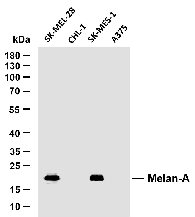
- Various whole cell lysates were separated by 10% SDS-PAGE, and the membrane was blotted with anti-Melan-A(ABT-Mart1) antibody. The HRP-conjugated Goat anti-Mouse IgG(H + L) antibody was used to detect the antibody. Lane 1: SK-MEL-28 Lane 2: CHL-1 Lane 3: SK-MES-1 Lane 4: A375 Predicted band size: 13kDa Observed band size: 18kDa


