Actin pan (ABT-ACTN) mouse mAb
- Catalog No.:YM6143
- Applications:WB; IHC;ELISA
- Reactivity:Human; Mouse (predicted: Rat; Bovin; Pig; Chick)
- Target:
- Actin pan
- Fields:
- >>Rap1 signaling pathway;>>Phagosome;>>Apoptosis;>>Hippo signaling pathway;>>Focal adhesion;>>Adherens junction;>>Tight junction;>>Platelet activation;>>Neutrophil extracellular trap formation;>>Leukocyte transendothelial migration;>>Thermogenesis;>>Regulation of actin cytoskeleton;>>Thyroid hormone signaling pathway;>>Oxytocin signaling pathway;>>Gastric acid secretion;>>Amyotrophic lateral sclerosis;>>Bacterial invasion of epithelial cells;>>Vibrio cholerae infection;>>Pathogenic Escherichia coli infection;>>Shigellosis;>>Salmonella infection;>>Yersinia infection;>>Influenza A;>>Proteoglycans in cancer;>>Hepatocellular carcinoma;>>Hypertrophic cardiomyopathy;>>Arrhythmogenic right ventricular cardiomyopathy;>>Dilated cardiomyopathy;>>Viral myocarditis;>>Fluid shear stress and atherosclerosis
- Gene Name:
- Actin-pan
- Protein Name:
- Actin pan
- Human Swiss Prot No:
- P60709/Q9BYX7/P63261
- Immunogen:
- Synthesized peptide derived from human Actin pan AA range: 300-377
- Specificity:
- This antibody detects endogenous levels of Actin pan. Heat-induced epitope retrieval (HIER) Citrate buffer of pH6.0 was highly recommended as antigen repair method in paraffin section
- Formulation:
- Liquid in PBS containing 50% glycerol, 0.5% BSA and 0.02% sodium azide.
- Source:
- Mouse, Monoclonal/IgG2a, Kappa
- Dilution:
- IHC 1:200-400,WB 1:500-2000, ELISA 1:5000-20000
- Purification:
- The antibody was affinity-purified from mouse ascites by affinity-chromatography using specific immunogen.
- Storage Stability:
- -15°C to -25°C/1 year(Do not lower than -25°C)
- Background:
- This gene encodes one of six different actin proteins. Actins are highly conserved proteins that are involved in cell motility, structure, and integrity. This actin is a major constituent of the contractile apparatus and one of the two nonmuscle cytoskeletal actins. [provided by RefSeq, Jul 2008],
- Function:
- disease:Defects in ACTB are a cause of dystonia juvenile-onset (DYTJ) [MIM:607371]. DYTJ is a form of dystonia with juvenile onset. Dystonia is defined by the presence of sustained involuntary muscle contraction, often leading to abnormal postures. DYTJ patients manifest progressive, generalized, dopa-unresponsive dystonia, developmental malformations and sensory hearing loss.,function:Actins are highly conserved proteins that are involved in various types of cell motility and are ubiquitously expressed in all eukaryotic cells.,miscellaneous:In vertebrates 3 main groups of actin isoforms, alpha, beta and gamma have been identified. The alpha actins are found in muscle tissues and are a major constituent of the contractile apparatus. The beta and gamma actins coexist in most cell types as components of the cytoskeleton and as mediators of internal cell motility.,similarity:Belongs to the
- Subcellular Location:
- Cytoplasm, cytoskeleton . Nucleus . Localized in cytoplasmic mRNP granules containing untranslated mRNAs. .
- Expression:
- B-cell lymphoma,Brain,Cajal-Retzius cell,Eye,Fetal brain cortex,Foreskin,Hepatocellular car
Ferritinophagy is involved in Bisphenol A-induced ferroptosis of renal tubular epithelial cells through the activation of the AMPK-mTOR-ULK1 pathway Food Chem Toxicol. 2022 May;163:112909. WB Mouse kidney tissue
- June 19-2018
- WESTERN IMMUNOBLOTTING PROTOCOL
- June 19-2018
- IMMUNOHISTOCHEMISTRY-PARAFFIN PROTOCOL
- June 19-2018
- IMMUNOFLUORESCENCE PROTOCOL
- September 08-2020
- FLOW-CYTOMEYRT-PROTOCOL
- May 20-2022
- Cell-Based ELISA│解您多样本WB检测之困扰
- July 13-2018
- CELL-BASED-ELISA-PROTOCOL-FOR-ACETYL-PROTEIN
- July 13-2018
- CELL-BASED-ELISA-PROTOCOL-FOR-PHOSPHO-PROTEIN
- July 13-2018
- Antibody-FAQs
- Products Images

- Human appendix tissue was stained with Anti-Actin pan (ABT-ACTN) Antibody

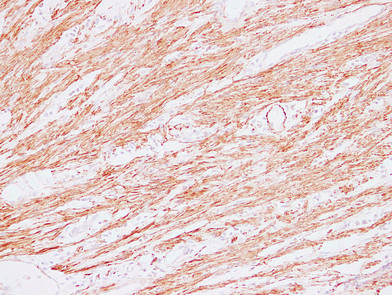
- Human cardiac muscle tissue was stained with Anti-Actin pan (ABT-ACTN) Antibody
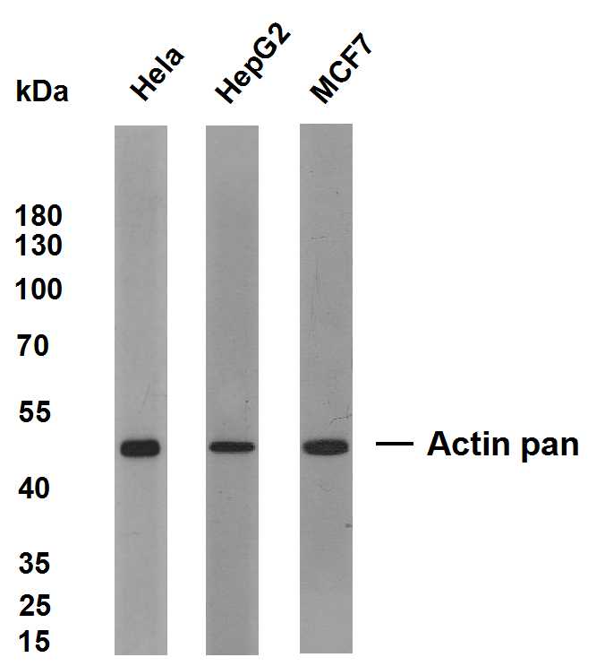
- Various whole cell lysates were separated by 10% SDS-PAGE, and the membrane was blotted with anti-Actin(ABT-ACTN) antibody. The HRP-conjugated Goat anti-Mouse IgG(H + L) antibody was used to detect the antibody. Lane 1: Hela Lane 2: HepG2 Lane 3: MCF7 Predicted band size: 42kDa Observed band size: 42kDa
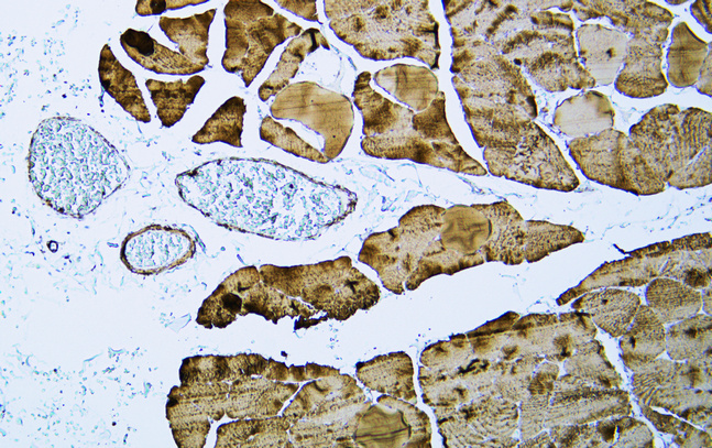
- Human skeletal muscle tissue was stained with Anti-Actin pan (ABT-ACTN) Antibody

- Human stomach tissue was stained with Anti-Actin pan (ABT-ACTN) Antibody
.jpg)
- Immunohistochemical analysis of paraffin-embedded Colon. 1, Antibody was diluted at 1:200(4° overnight). 2, Citric acid ,pH6.0 was used for antigen retrieval. 3,Secondary antibody was diluted at 1:200(room temperature, 30min).
.jpg)
- Immunohistochemical analysis of paraffin-embedded Colon. 1, Antibody was diluted at 1:200(4° overnight). 2, Citric acid ,pH6.0 was used for antigen retrieval. 3,Secondary antibody was diluted at 1:200(room temperature, 30min).
.jpg)
- Immunohistochemical analysis of paraffin-embedded Colon. 1, Antibody was diluted at 1:200(4° overnight). 2, Citric acid ,pH6.0 was used for antigen retrieval. 3,Secondary antibody was diluted at 1:200(room temperature, 30min).
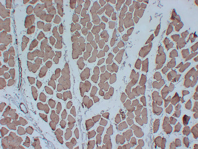
- Immunohistochemical analysis of paraffin-embedded Skeletal muscle. 1, Antibody was diluted at 1:200(4° overnight). 2, Citric acid ,pH6.0 was used for antigen retrieval. 3,Secondary antibody was diluted at 1:200(room temperature, 30min).
- Immunohistochemical analysis of paraffin-embedded Colon. 1, Antibody was diluted at 1:200(4° overnight). 2, Citric acid ,pH6.0 was used for antigen retrieval. 3,Secondary antibody was diluted at 1:200(room temperature, 30min).
_wb.jpg)
- Western blot analysis of Actin-panAntibody at 1:1000 dilution.
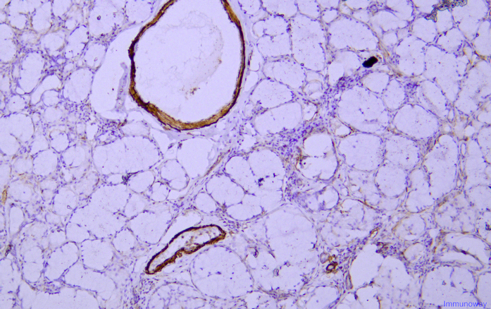
- Immunohistochemical analysis of paraffin-embedded human Salivary gland tonsils Antibody was diluted at 1:200(4° overnight).

- Immunohistochemical analysis of paraffin-embedded human Salivary gland tonsils Antibody was diluted at 1:200(4° overnight).


