Bax (PTR1159) mouse mAb
- Catalog No.:YM3619
- Applications:WB;ELISA
- Reactivity:Human;
- Target:
- Bax
- Fields:
- >>EGFR tyrosine kinase inhibitor resistance;>>Endocrine resistance;>>Platinum drug resistance;>>Sphingolipid signaling pathway;>>p53 signaling pathway;>>Protein processing in endoplasmic reticulum;>>Apoptosis;>>Longevity regulating pathway;>>Apoptosis - multiple species;>>Necroptosis;>>Neurotrophin signaling pathway;>>Non-alcoholic fatty liver disease;>>AGE-RAGE signaling pathway in diabetic complications;>>Parkinson disease;>>Amyotrophic lateral sclerosis;>>Huntington disease;>>Prion disease;>>Pathways of neurodegeneration - multiple diseases;>>Pathogenic Escherichia coli infection;>>Shigellosis;>>Salmonella infection;>>Tuberculosis;>>Hepatitis C;>>Hepatitis B;>>Measles;>>Human cytomegalovirus infection;>>Influenza A;>>Human papillomavirus infection;>>Human T-cell leukemia virus 1 infection;>>Kaposi sarcoma-associated herpesvirus infection;>>Herpes simplex virus 1 infection;>>Epstein-Barr virus infection;>>Human immunodeficiency virus 1 infection;>>Pathways in cancer;>>Transcriptional
- Gene Name:
- BAX
- Protein Name:
- BAX
- Human Gene Id:
- 581
- Human Swiss Prot No:
- Q07812
- Immunogen:
- Synthesized peptide derived from human Bax AA range: 50-150
- Specificity:
- This antibody detects endogenous levels of BAX.
- Formulation:
- PBS, 50% glycerol, 0.05% Proclin 300, 0.05%BSA
- Source:
- Monoclonal, Mouse IgG1, Kappa
- Dilution:
- WB 1:500-1000, ELISA 1:5000-20000
- Purification:
- Protein G
- Storage Stability:
- -15°C to -25°C/1 year(Do not lower than -25°C)
- Other Name:
- BAX;BCL2L4
- Molecular Weight(Da):
- 21kD
- Observed Band(KD):
- 21kD
- Background:
- The protein encoded by BAX (BCL2 associated X, apoptosis regulator) belongs to the BCL2 protein family. BCL2 family members form hetero- or homodimers and act as anti- or pro-apoptotic regulators that are involved in a wide variety of cellular activities. This protein forms a heterodimer with BCL2, and functions as an apoptotic activator. This protein is reported to interact with, and increase the opening of, the mitochondrial voltage-dependent anion channel (VDAC), which leads to the loss in membrane potential and the release of cytochrome c. The expression of this gene is regulated by the tumor suppressor P53 and has been shown to be involved in P53-mediated apoptosis. Multiple alternatively spliced transcript variants, which encode different isoforms, have been reported for BAX.
- Function:
- disease:Defects in BAX are found in some cell lines from hematopoietic malignancies as T-cell acute lymphoblastic leukemia, Burkitt lymphoma, and plasmacytoma.,domain:Intact BH3 motif is required by BIK, BID, BAK, BAD and BAX for their pro-apoptotic activity and for their interaction with anti-apoptotic members of the Bcl-2 family.,function:Accelerates programmed cell death by binding to, and antagonizing the apoptosis repressor BCL2 or its adenovirus homolog E1B 19k protein. Induces the release of cytochrome c, activation of CASP3, and thereby apoptosis.,similarity:Belongs to the Bcl-2 family.,subcellular location:Colocalizes with 14-3-3 proteins in the cytoplasm. Under stress conditions, redistributes to the mitochondrion membrane through the release from JNK-phosphorylated 14-3-3 proteins.,subunit:Homodimer. Forms heterodimers with BCL2, E1B 19K protein, BCL2L1 isoform Bcl-X(L), MCL1
- Subcellular Location:
- [Isoform Alpha]: Mitochondrion outer membrane ; Single-pass membrane protein . Cytoplasm . Colocalizes with 14-3-3 proteins in the cytoplasm. Under stress conditions, undergoes a conformation change that causes release from JNK-phosphorylated 14-3-3 proteins and translocation to the mitochondrion membrane. Upon Sendai virus infection, recruited to the mitochondrion through interaction with IRF3 (PubMed:25609812). .; [Isoform Beta]: Cytoplasm.; [Isoform Gamma]: Cytoplasm.; [Isoform Delta]: Cytoplasm .
- Expression:
- Expressed in a wide variety of tissues. Isoform Psi is found in glial tumors. Isoform Alpha is expressed in spleen, breast, ovary, testis, colon and brain, and at low levels in skin and lung. Isoform Sigma is expressed in spleen, breast, ovary, testis, lung, colon, brain and at low levels in skin. Isoform Alpha and isoform Sigma are expressed in pro-myelocytic leukemia, histiocytic lymphoma, Burkitt's lymphoma, T-cell lymphoma, lymphoblastic leukemia, breast adenocarcinoma, ovary adenocarcinoma, prostate carcinoma, prostate adenocarcinoma, lung carcinoma, epidermoid carcinoma, small cell lung carcinoma and colon adenocarcinoma cell lines.
Genetically engineered magnetic nanocages for cancer magneto-catalytic theranostics. Nature Communications Nat Commun. 2020 Oct;11(1):1-11 WB Human A549 cell
A preparation of Ginkgo biloba L. leaves extract inhibits the apoptosis of hippocampal neurons in post-stroke mice via regulating the expression of Bax/Bcl-2 and Caspase-3. JOURNAL OF ETHNOPHARMACOLOGY J Ethnopharmacol. 2021 Nov;280:114481 IHC,IF,WB Mouse 1:110,1:1000 Hippocampus Hippocampal cell line HT-22
The Effect of SPTLC2 on Promoting Neuronal Apoptosis is Alleviated by MiR-124-3p Through TLR4 Signalling Pathway. NEUROCHEMICAL RESEARCH Neurochem Res. 2019 Sep;44(9):2113-2122 WB Mouse 1:2000 cortical neurons
Emodin suppresses the migration and invasion of melanoma cells. BIOLOGICAL & PHARMACEUTICAL BULLETIN 2021 Mar 17 WB Mouse,Human 1 : 500 B16F10 Melanoma Cells, A375 Melanoma Cells
HN1L is essential for cell growth and survival during nucleopolyhedrovirus infection in silkworm, Bombyx mori. PLoS One Plos One. 2019 May;14(5):e0216719 WB Silkworm 1:1000 BmN cell
Mesenchymal stem cell-derived extracellular vesicles promote apoptosis in RSC96 Schwann cells through the activation of the ERK pathway. International Journal of Clinical and Experimental Pathology Int J Clin Exp Patho. 2018; 11(11): 5157–5170 WB Rat RSC96 cell
Suppression of Cellular Proliferation and Invasion by HMGB1 Knockdown in Bladder Urothelial Carcinoma Cells. ONCOLOGY RESEARCH Oncol Res. 2015; 22(5-6): 235–245 WB Human BIU-87 cell, T24 cell
Establishment and Mechanism Study of a Primary Ovarian Insufficiency Mouse Model Using Lipopolysaccharide. Analytical Cellular Pathology Anal Cell Pathol. 2021;2021:1781532 WB Mouse 1:1000 Ovarian tissue
Chlamydia psittaci inclusion membrane protein CPSIT_0842 induces macrophage apoptosis through MAPK/ERK-mediated autophagy INTERNATIONAL JOURNAL OF BIOCHEMISTRY & CELL BIOLOGY Yimou Wu WB Human human monocytic leukemia cell line THP-1 cell
L-Theanine delays D-galactose-induced senescence by regulating the cell cycle and inhibiting apoptosis in rat intestinal cells JOURNAL OF THE SCIENCE OF FOOD AND AGRICULTURE Wei Xu WB Rat 1:2000 IEC-6 cell
- June 19-2018
- WESTERN IMMUNOBLOTTING PROTOCOL
- June 19-2018
- IMMUNOHISTOCHEMISTRY-PARAFFIN PROTOCOL
- June 19-2018
- IMMUNOFLUORESCENCE PROTOCOL
- September 08-2020
- FLOW-CYTOMEYRT-PROTOCOL
- May 20-2022
- Cell-Based ELISA│解您多样本WB检测之困扰
- July 13-2018
- CELL-BASED-ELISA-PROTOCOL-FOR-ACETYL-PROTEIN
- July 13-2018
- CELL-BASED-ELISA-PROTOCOL-FOR-PHOSPHO-PROTEIN
- July 13-2018
- Antibody-FAQs
- Products Images
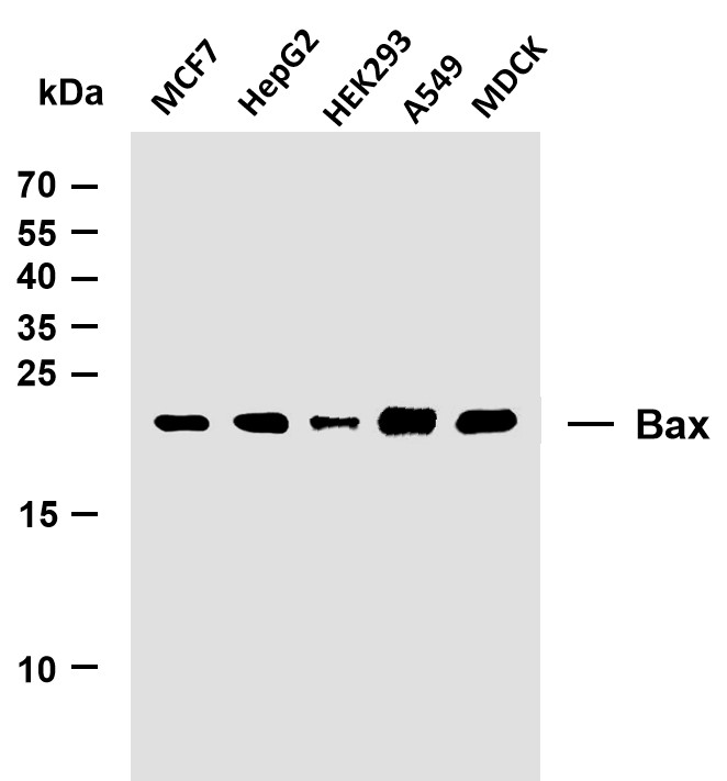
- Various whole cell lysates were separated by 15% SDS-PAGE, and the membrane was blotted with anti-Bax (PTR1159) antibody. The HRP-conjugated Goat anti-Mouse IgG(H + L) antibody was used to detect the antibody. Lane 1: MCF7 Lane 2: HepG2 Lane 3: HEK293 Lane 4: A549 Lane 5: MDCK Predicted band size: 21kDa Observed band size: 21kDa
.jpg)
- Zhang, Y., Wang, X., Chu, C. et al. Genetically engineered magnetic nanocages for cancer magneto-catalytic theranostics. Nat Commun 11, 5421 (2020).
- Chen, Puxiang, et al. "Long noncoding RNA LINC00152 promotes cell proliferation through competitively binding endogenous miR‐125b with MCL‐1 by regulating mitochondrial apoptosis pathways in ovarian cancer." Cancer medicine 7.9 (2018): 4530-4541.
- Wang, Yupei, et al. "Selective ATP hydrolysis inhibition in F1Fo ATP synthase enhances radiosensitivity in non-small-cell lung cancer cells (A549)." Oncotarget 8.32 (2017): 53602.
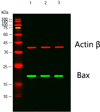
- Western blot analysis of lysates from 1) Hela Cell Lysate, 2) C2C12 Cell Lysate, 3) PC12 Cell Lysate cells, (Green) primary antibody was diluted at 1:1000, 4°over night,Dylight 800 secondary antibody(Immunoway:RS23910)was diluted at 1:10000, 37° 1hour. (Red) Actin β Polyclonal Antibody (Immunoway:YT0099) antibody was diluted at 1:5000 as loading control, 4° over night,Dylight 680 secondary antibody(Immunoway:RS23720)was diluted at 1:10000, 37° 1hour.
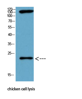
- Western Blot analysis of chicken cell lysis using Antibody diluted at 1:1000


