Histone H3 (Tri Methyl Lys79) Monoclonal Antibody(3G3)
- Catalog No.:YM3091
- Applications:WB;IHC;IF;IP
- Reactivity:Human;Mouse;Rat
- Target:
- Histone H3
- Fields:
- >>Neutrophil extracellular trap formation;>>Alcoholism;>>Shigellosis;>>Transcriptional misregulation in cancer;>>Systemic lupus erythematosus
- Gene Name:
- HIST1H3A/HIST1H3B/HIST1H3C/HIST1H3D/HIST1H3E/HIST1H3F/HIST1H3G/HIST1H3H/HIST1H3I/HIST1H3J/HIST2H3A/HIST2H3C/HIST2H3D/H3F3A/H3F3B
- Protein Name:
- Histone H3.1/Histone H3.2/Histone H3.3
- Human Gene Id:
- 8350/8351/8352/8353/8354/8355/8356/8357/8358/8968
- Human Swiss Prot No:
- P68431/Q71DI3/P84243
- Mouse Gene Id:
- 319152/15077/15078
- Rat Gene Id:
- 291159/100361558
- Rat Swiss Prot No:
- Q6LED0/P84245
- Immunogen:
- Synthetic Peptide of Histone H3 (Tri Methyl Lys79)
- Specificity:
- The antibody detects endogenous Histone H3 (tri methyl K79) protein.
- Formulation:
- PBS, pH 7.4, containing 0.5%BSA, 0.02% sodium azide as Preservative and 50% Glycerol.
- Source:
- Monoclonal, Mouse
- Dilution:
- WB 1:500-2000 IP:1:200 IF 1:200 IHC 1:50-300
- Purification:
- The antibody was affinity-purified from mouse ascites by affinity-chromatography using specific immunogen.
- Storage Stability:
- -15°C to -25°C/1 year(Do not lower than -25°C)
- Other Name:
- H3K79ME3;HIST1H3A;H3FA;HIST1H3B;H3FL;HIST1H3C;H3FC;HIST1H3D;H3FB;HIST1H3E;H3FD;HIST1H3F;H3FI;HIST1H3G;H3FH;HIST1H3H;H3FK;HIST1H3I;H3FF;HIST1H3J;H3FJ;Histone H3.1;Histone H3/a;Histone H3/b;Histone H3/c;Histone H3/d;Histone H3/f;Histone H3/h;Histone H3/i;Histone H3/j;Histone H3/k;Histone H3/l;HIST2H3A;HIST2H3C;H3F2;H3FM;HIST2H3D;Histone H3.2;Histone H3/m;Histone H3/o;H3F3A;H3.3A;H3F3;PP781;H3F3B;H3.3B;Histone H3.3
- Observed Band(KD):
- 15kD
- Background:
- Histones are basic nuclear proteins that are responsible for the nucleosome structure of the chromosomal fiber in eukaryotes. This structure consists of approximately 146 bp of DNA wrapped around a nucleosome, an octamer composed of pairs of each of the four core histones (H2A, H2B, H3, and H4). The chromatin fiber is further compacted through the interaction of a linker histone, H1, with the DNA between the nucleosomes to form higher order chromatin structures. This gene is intronless and encodes a replication-dependent histone that is a member of the histone H3 family. Transcripts from this gene lack polyA tails; instead, they contain a palindromic termination element. This gene is found in the large histone gene cluster on chromosome 6p22-p21.3. [provided by RefSeq, Aug 2015],
- Function:
- caution:Was originally (PubMed:2587222) thought to originate from mouse.,developmental stage:Expressed during S phase, then expression strongly decreases as cell division slows down during the process of differentiation.,function:Core component of nucleosome. Nucleosomes wrap and compact DNA into chromatin, limiting DNA accessibility to the cellular machineries which require DNA as a template. Histones thereby play a central role in transcription regulation, DNA repair, DNA replication and chromosomal stability. DNA accessibility is regulated via a complex set of post-translational modifications of histones, also called histone code, and nucleosome remodeling.,mass spectrometry:Monoisotopic with N-acetylserine PubMed:16457589,miscellaneous:This histone is only present in mammals and is enriched in acetylation of Lys-15 and dimethylation of Lys-10 (H3K9me2).,PTM:Acetylation is generally l
- Subcellular Location:
- Nucleus. Chromosome.
- Expression:
- Blood,Epithelium,Kidney,Lung,Ovary,Spleen,Uterus,
In Colorectal Cancer Cells With Mutant KRAS, SLC25A22-Mediated Glutaminolysis Reduces DNA Demethylation to Increase WNT Signaling, Stemness, and Drug Resistance. GASTROENTEROLOGY 2020 Aug 16 WB Human DLD1 cell, HCT116 cell
The elevated transcription of ADAM19 by the oncohistone H2BE76K contributes to oncogenic properties in breast cancer. JOURNAL OF BIOLOGICAL CHEMISTRY 2021 Jan;296: WB Human 293T cell,MDA-MB-231 cell
The elevated transcription of ADAM19 by the oncohistone H2BE76K contributes to oncogenic properties in breast cancer. JOURNAL OF BIOLOGICAL CHEMISTRY 2021 Jan;296: WB Human 293T cell,MDA-MB-231 cell
- June 19-2018
- WESTERN IMMUNOBLOTTING PROTOCOL
- June 19-2018
- IMMUNOHISTOCHEMISTRY-PARAFFIN PROTOCOL
- June 19-2018
- IMMUNOFLUORESCENCE PROTOCOL
- September 08-2020
- FLOW-CYTOMEYRT-PROTOCOL
- May 20-2022
- Cell-Based ELISA│解您多样本WB检测之困扰
- July 13-2018
- CELL-BASED-ELISA-PROTOCOL-FOR-ACETYL-PROTEIN
- July 13-2018
- CELL-BASED-ELISA-PROTOCOL-FOR-PHOSPHO-PROTEIN
- July 13-2018
- Antibody-FAQs
- Products Images
.jpg)
- Kang, Tze Zhen Evangeline, et al. "The elevated transcription of ADAM19 by the oncohistone H2BE76K contributes to oncogenic properties in breast cancer." Journal of Biological Chemistry 296 (2021).
.jpg)
- Wong, Chi Chun, et al. "In Colorectal Cancer Cells With Mutant KRAS, SLC25A22-Mediated Glutaminolysis Reduces DNA Demethylation to Increase WNT Signaling, Stemness, and Drug Resistance." Gastroenterology 159.6 (2020): 2163-2180.
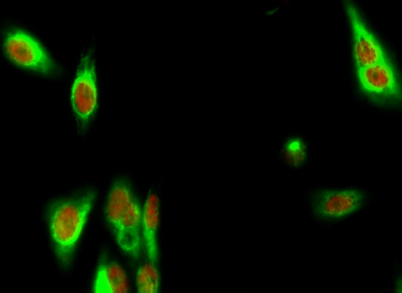
- Immunofluorescence analysis of Hela cell. 1,Bek Polyclonal Antibody(green) was diluted at 1:200(4° overnight). (red) was diluted at 1:200(4° overnight). 2, Goat Anti Rabbit Alexa Fluor 488 Catalog:RS3211 was diluted at 1:1000(room temperature, 50min). Goat Anti Mouse Alexa Fluor 594 Catalog:RS3608 was diluted at 1:1000(room temperature, 50min).
mono-ihc-human-uterus.jpg)
- Immunohistochemical analysis of paraffin-embedded Human-uterus tissue. 1,Histone H3 (Tri Methyl Lys79) Monoclonal Antibody(3G3) was diluted at 1:200(4°C,overnight). 2, Sodium citrate pH 6.0 was used for antibody retrieval(>98°C,20min). 3,Secondary antibody was diluted at 1:200(room tempeRature, 30min). Negative control was used by secondary antibody only.
mono-ihc-rat-heart.jpg)
- Immunohistochemical analysis of paraffin-embedded Rat-heart tissue. 1,Histone H3 (Tri Methyl Lys79) Monoclonal Antibody(3G3) was diluted at 1:200(4°C,overnight). 2, Sodium citrate pH 6.0 was used for antibody retrieval(>98°C,20min). 3,Secondary antibody was diluted at 1:200(room tempeRature, 30min). Negative control was used by secondary antibody only.
mono-ihc-mouse-heart.jpg)
- Immunohistochemical analysis of paraffin-embedded Mouse-heart tissue. 1,Histone H3 (Tri Methyl Lys79) Monoclonal Antibody(3G3) was diluted at 1:200(4°C,overnight). 2, Sodium citrate pH 6.0 was used for antibody retrieval(>98°C,20min). 3,Secondary antibody was diluted at 1:200(room tempeRature, 30min). Negative control was used by secondary antibody only.
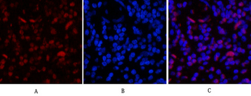
- Immunofluorescence analysis of Human-appendix tissue. 1,Histone H3 (Tri Methyl Lys79) Monoclonal Antibody(3G3)(red) was diluted at 1:200(4°C,overnight). 2, Cy3 labled Secondary antibody was diluted at 1:300(room temperature, 50min).3, Picture B: DAPI(blue) 10min. Picture A:Target. Picture B: DAPI. Picture C: merge of A+B

- Immunofluorescence analysis of Rat-liver tissue. 1,Histone H3 (Tri Methyl Lys79) Monoclonal Antibody(3G3)(red) was diluted at 1:200(4°C,overnight). 2, Cy3 labled Secondary antibody was diluted at 1:300(room temperature, 50min).3, Picture B: DAPI(blue) 10min. Picture A:Target. Picture B: DAPI. Picture C: merge of A+B

- Western blot analysis of Hela, diluted at 1) 1:2000 2) 1:5000 cells nucleus extracted by Minute TM Cytoplasmic and Nuclear Fractionation kit (SC-003,Inventbiotech,MN,USA).

- 1) Input: Hela Cell Lysate 2) IP product: IP dilute 1:200


