GFAP Monoclonal Antibody(5C8)
- Catalog No.:YM3059
- Applications:WB;IHC;IF;
- Reactivity:Human;Rat;Mouse
- Target:
- GFAP
- Fields:
- >>JAK-STAT signaling pathway
- Gene Name:
- GFAP
- Protein Name:
- Glial fibrillary acidic protein
- Human Gene Id:
- 2670
- Human Swiss Prot No:
- P14136
- Mouse Gene Id:
- 14580
- Mouse Swiss Prot No:
- P03995
- Rat Gene Id:
- 24387
- Rat Swiss Prot No:
- P47819
- Immunogen:
- Synthetic Peptide of GFAP
- Specificity:
- The antibody detects endogenous GFAP proteins.
- Formulation:
- PBS, pH 7.4, containing 0.5%BSA, 0.02% sodium azide as Preservative and 50% Glycerol.
- Source:
- Monoclonal, Mouse
- Dilution:
- WB 1:2000-5000 IF 1:200 IHC 1:50-300
- Purification:
- The antibody was affinity-purified from mouse ascites by affinity-chromatography using specific immunogen.
- Storage Stability:
- -15°C to -25°C/1 year(Do not lower than -25°C)
- Other Name:
- GFAP;Glial fibrillary acidic protein;GFAP
- Observed Band(KD):
- 45kD
- Background:
- This gene encodes one of the major intermediate filament proteins of mature astrocytes. It is used as a marker to distinguish astrocytes from other glial cells during development. Mutations in this gene cause Alexander disease, a rare disorder of astrocytes in the central nervous system. Alternative splicing results in multiple transcript variants encoding distinct isoforms. [provided by RefSeq, Oct 2008],
- Function:
- alternative products:Isoforms differ in the C-terminal region which is encoded by alternative exons,disease:Defects in GFAP are a cause of Alexander disease (ALEXD) [MIM:203450]. Alexander disease is a rare disorder of the central nervous system. It is a progressive leukoencephalopathy whose hallmark is the widespread accumulation of Rosenthal fibers which are cytoplasmic inclusions in astrocytes. The most common form affects infants and young children, and is characterized by progressive failure of central myelination, usually leading to death usually within the first decade. Infants with Alexander disease develop a leukoencephalopathy with macrocephaly, seizures, and psychomotor retardation. Patients with juvenile or adult forms typically experience ataxia, bulbar signs and spasticity, and a more slowly progressive course.,function:GFAP, a class-III intermediate filament, is a cell-spe
- Subcellular Location:
- Cytoplasm . Associated with intermediate filaments. .
- Expression:
- Expressed in cells lacking fibronectin.
Elemene Emulsion Injection Administration Reduces Neuropathic Pain by Inhibiting Astrocytic NDRG2 Expression within Spinal Dorsal Horn. Chinese Journal of Integrative Medicine Chin J Integr Med. 2021 Dec;27(12):912-918 WB Rat 1:1000 Lumbar spinal cord
Effects of altitude changes on mild‑to‑moderate closed‑head injury in rats following acute high‑altitude exposure. Experimental and Therapeutic Medicine 2018 Nov 27 IHC Rat Brain tissues
平昊达, et al. "慢性脑低灌注大鼠焦虑样行为与海马区域炎性细胞因子水平的相关性分析." 收藏 3 (2020).
The analgesic effects of β-elemene in rats with neuropathic pain by inhibition of spinal astrocytic ERK activation: Molecular Pain Jin Zheng WB Rat
Bu Shen Yi Sui Capsule Promotes Myelin Repair by Modulating the Transformation of A1/A2 Reactive Astrocytes In Vivo and In Vitro Oxidative Medicine and Cellular Longevity Lei Wang IHC, IF, WB Mouse, Rat
- June 19-2018
- WESTERN IMMUNOBLOTTING PROTOCOL
- June 19-2018
- IMMUNOHISTOCHEMISTRY-PARAFFIN PROTOCOL
- June 19-2018
- IMMUNOFLUORESCENCE PROTOCOL
- September 08-2020
- FLOW-CYTOMEYRT-PROTOCOL
- May 20-2022
- Cell-Based ELISA│解您多样本WB检测之困扰
- July 13-2018
- CELL-BASED-ELISA-PROTOCOL-FOR-ACETYL-PROTEIN
- July 13-2018
- CELL-BASED-ELISA-PROTOCOL-FOR-PHOSPHO-PROTEIN
- July 13-2018
- Antibody-FAQs
- Products Images
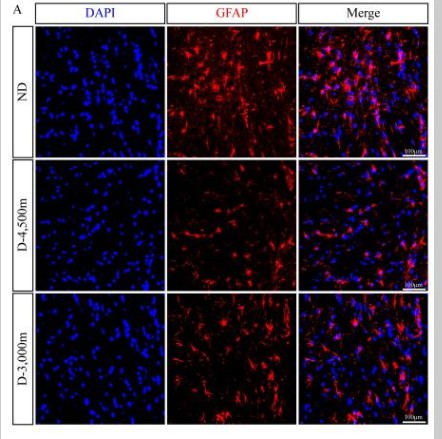
- Wang, Hao, et al. "Effects of altitude changes on mild‑to‑moderate closed‑head injury in rats following acute high‑altitude exposure." Experimental and therapeutic medicine 17.1 (2019): 847-856.

- Immunofluorescence analysis of Hela cell. 1,AR Polyclonal Antibody(red) was diluted at 1:200(4° overnight). GFAP Monoclonal Antibody(5C8)(green) was diluted at 1:200(4° overnight). 2, Goat Anti Rabbit Alexa Fluor 594 Catalog:RS3611 was diluted at 1:1000(room temperature, 50min). Goat Anti Mouse Alexa Fluor 488 Catalog:RS3208 was diluted at 1:1000(room temperature, 50min).
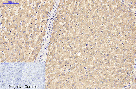
- Immunohistochemical analysis of paraffin-embedded Human-liver tissue. 1,GFAP Monoclonal Antibody(5C8) was diluted at 1:200(4°C,overnight). 2, Sodium citrate pH 6.0 was used for antibody retrieval(>98°C,20min). 3,Secondary antibody was diluted at 1:200(room tempeRature, 30min). Negative control was used by secondary antibody only.
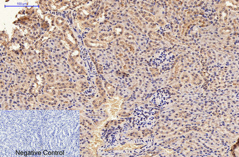
- Immunohistochemical analysis of paraffin-embedded Mouse-kidney tissue. 1,GFAP Monoclonal Antibody(5C8) was diluted at 1:200(4°C,overnight). 2, Sodium citrate pH 6.0 was used for antibody retrieval(>98°C,20min). 3,Secondary antibody was diluted at 1:200(room tempeRature, 30min). Negative control was used by secondary antibody only.
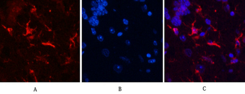
- Immunofluorescence analysis of Mouse-brain tissue. 1,GFAP Monoclonal Antibody(5C8)(red) was diluted at 1:200(4°C,overnight). 2, Cy3 labled Secondary antibody was diluted at 1:300(room temperature, 50min).3, Picture B: DAPI(blue) 10min. Picture A:Target. Picture B: DAPI. Picture C: merge of A+B
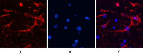
- Immunofluorescence analysis of Rat-brain tissue. 1,GFAP Monoclonal Antibody(5C8)(red) was diluted at 1:200(4°C,overnight). 2, Cy3 labled Secondary antibody was diluted at 1:300(room temperature, 50min).3, Picture B: DAPI(blue) 10min. Picture A:Target. Picture B: DAPI. Picture C: merge of A+B
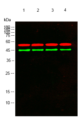
- Western blot analysis of lysates from 1) Rat Brain Tissue, 2)HeLa , 3)A431, 4) PC12 cells, (Green) primary antibody was diluted at 1:1000, 4°over night, secondary antibody(cat:RS23910)was diluted at 1:10000, 37° 1hour. (Red) Tubulin β Polyclonal Antibody (cat:YT4780) antibody was diluted at 1:5000 as loading control, 4° over night,secondary antibody(cat:RS23720)was diluted at 1:10000, 37° 1hour.


