p53 Monoclonal Antibody(6C4)
- Catalog No.:YM3052
- Applications:WB;IHC;IF
- Reactivity:Human;Mouse
- Target:
- p53
- Fields:
- >>Endocrine resistance;>>Platinum drug resistance;>>MAPK signaling pathway;>>Sphingolipid signaling pathway;>>Cell cycle;>>p53 signaling pathway;>>Mitophagy - animal;>>PI3K-Akt signaling pathway;>>Apoptosis;>>Longevity regulating pathway;>>Ferroptosis;>>Cellular senescence;>>Wnt signaling pathway;>>Neurotrophin signaling pathway;>>Thyroid hormone signaling pathway;>>Parkinson disease;>>Amyotrophic lateral sclerosis;>>Huntington disease;>>Shigellosis;>>Hepatitis C;>>Hepatitis B;>>Measles;>>Human cytomegalovirus infection;>>Human papillomavirus infection;>>Human T-cell leukemia virus 1 infection;>>Kaposi sarcoma-associated herpesvirus infection;>>Herpes simplex virus 1 infection;>>Epstein-Barr virus infection;>>Pathways in cancer;>>Transcriptional misregulation in cancer;>>Viral carcinogenesis;>>Proteoglycans in cancer;>>MicroRNAs in cancer;>>Colorectal cancer;>>Pancreatic cancer;>>Endometrial cancer;>>Glioma;>>Prostate cancer;>>Thyroid cancer;>>Basal cell carcinoma;>>Melanoma;>>Bladder
- Gene Name:
- TP53
- Protein Name:
- Cellular tumor antigen p53
- Human Gene Id:
- 7157
- Human Swiss Prot No:
- P04637
- Mouse Gene Id:
- 22059
- Mouse Swiss Prot No:
- P02340
- Rat Gene Id:
- 24842
- Rat Swiss Prot No:
- P10361
- Immunogen:
- Synthetic Peptide of p53。AA 10-59
- Specificity:
- The antibody detects endogenous p53 proteins.
- Formulation:
- PBS, pH 7.4, containing 0.5%BSA, 0.02% sodium azide as Preservative and 50% Glycerol.
- Source:
- Monoclonal, Mouse
- Dilution:
- WB 1:2000 IHC 1:200 IF 1:100-200
- Purification:
- The antibody was affinity-purified from mouse ascites by affinity-chromatography using specific immunogen.
- Storage Stability:
- -15°C to -25°C/1 year(Do not lower than -25°C)
- Other Name:
- Antigen NY-CO-13;BCC7;Cellular tumor antigen p53;FLJ92943;LFS1;Mutant tumor protein 53;p53;p53 tumor suppressor;P53_HUMAN;Phosphoprotein p53;Tp53;Transformation related protein 53;TRP53;tumor antigen p55;Tumor protein 53;Tumor protein p53;Tumor suppressor p53;
- Observed Band(KD):
- 53kD
- Background:
- tumor protein p53(TP53) Homo sapiens This gene encodes a tumor suppressor protein containing transcriptional activation, DNA binding, and oligomerization domains. The encoded protein responds to diverse cellular stresses to regulate expression of target genes, thereby inducing cell cycle arrest, apoptosis, senescence, DNA repair, or changes in metabolism. Mutations in this gene are associated with a variety of human cancers, including hereditary cancers such as Li-Fraumeni syndrome. Alternative splicing of this gene and the use of alternate promoters result in multiple transcript variants and isoforms. Additional isoforms have also been shown to result from the use of alternate translation initiation codons (PMIDs: 12032546, 20937277). [provided by RefSeq, Feb 2013],
- Function:
- cofactor:Binds 1 zinc ion per subunit.,disease:Defects in TP53 are a cause of choroid plexus papilloma [MIM:260500]. Choroid plexus papilloma is a slow-growing benign tumor of the choroid plexus that often invades the leptomeninges. In children it is usually in a lateral ventricle but in adults it is more often in the fourth ventricle. Hydrocephalus is common, either from obstruction or from tumor secretion of cerebrospinal fluid. If it undergoes malignant transformation it is called a choroid plexus carcinoma. Primary choroid plexus tumors are rare and usually occur in early childhood.,disease:Defects in TP53 are a cause of Li-Fraumeni syndrome (LFS) [MIM:151623]. LFS is an autosomal dominant familial cancer syndrome that in its classic form is defined by the existence of a proband affected by a sarcoma before 45 years with a first degree relative affected by any tumor before 45 years a
- Subcellular Location:
- Cytoplasm . Nucleus . Nucleus, PML body . Endoplasmic reticulum . Mitochondrion matrix . Cytoplasm, cytoskeleton, microtubule organizing center, centrosome . Recruited into PML bodies together with CHEK2 (PubMed:12810724). Translocates to mitochondria upon oxidative stress (PubMed:22726440). Translocates to mitochondria in response to mitomycin C treatment (PubMed:27323408). .; [Isoform 1]: Nucleus . Cytoplasm. Predominantly nuclear but localizes to the cytoplasm when expressed with isoform 4.; [Isoform 2]: Nucleus. Cytoplasm. Localized mainly in the nucleus with minor staining in the cytoplasm.; [Isoform 3]: Nucleus. Cytoplasm. Localized in the nucleus in most cells but found in the cytoplasm in some cells.; [Isoform 4]: Nucleus. Cytoplasm. Predominantly nuclear but translocates to the cy
- Expression:
- Ubiquitous. Isoforms are expressed in a wide range of normal tissues but in a tissue-dependent manner. Isoform 2 is expressed in most normal tissues but is not detected in brain, lung, prostate, muscle, fetal brain, spinal cord and fetal liver. Isoform 3 is expressed in most normal tissues but is not detected in lung, spleen, testis, fetal brain, spinal cord and fetal liver. Isoform 7 is expressed in most normal tissues but is not detected in prostate, uterus, skeletal muscle and breast. Isoform 8 is detected only in colon, bone marrow, testis, fetal brain and intestine. Isoform 9 is expressed in most normal tissues but is not detected in brain, heart, lung, fetal liver, salivary gland, breast or intestine.
Chidamide Combined With Doxorubicin Induced p53-Driven Cell Cycle Arrest and Cell Apoptosis Reverse Multidrug Resistance of Breast Cancer. Frontiers in Oncology Front Oncol. 2021 Mar;0:344 WB Human 1:2000 CALDOX cell,MCF-7 cell,A02 cell
Tang, Junxia, et al. "TEM1 表达与胃癌患者新辅助化疗疗效相关性研究." 中国肿瘤临床 46.4 (2019): 173-177.
- June 19-2018
- WESTERN IMMUNOBLOTTING PROTOCOL
- June 19-2018
- IMMUNOHISTOCHEMISTRY-PARAFFIN PROTOCOL
- June 19-2018
- IMMUNOFLUORESCENCE PROTOCOL
- September 08-2020
- FLOW-CYTOMEYRT-PROTOCOL
- May 20-2022
- Cell-Based ELISA│解您多样本WB检测之困扰
- July 13-2018
- CELL-BASED-ELISA-PROTOCOL-FOR-ACETYL-PROTEIN
- July 13-2018
- CELL-BASED-ELISA-PROTOCOL-FOR-PHOSPHO-PROTEIN
- July 13-2018
- Antibody-FAQs
- Products Images

- P53 knockout and wild-type HEK93 whole cell lysates were separated by 4-20% SDS-PAGE, and the membrane was blotted with anti-P53 and anti-Actin antibody. The HRP-conjugated anti-Mouse IgG antibody was used to detect the antibody. Lane1: Wild type HEK293 whole cell lysate, 20ug; Lane2: P53 knockout HEK293 whole cell lysate, 20ug; Predicted band size: 53 kDa Observed band size: 53 kDa

- IHC staining of Human colon cancer tissue paraffin-embedded, diluted at 1:200.
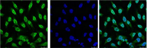
- IF analysis of Hela with antibody (Left) and DAPI (Right) diluted at 1:100.
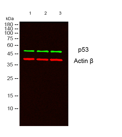
- Western blot analysis of lysates from 1) 293T, 2) HEK293, 3) A431 cells, (Green) primary antibody was diluted at 1:1000, 4°over night, secondary antibody(cat:RS23910)was diluted at 1:10000, 37° 1hour. (Red) Actin β Polyclonal Antibody (cat:YT0099) antibody was diluted at 1:5000 as loading control, 4° over night,secondary antibody(cat:RS23720)was diluted at 1:10000, 37° 1hour.
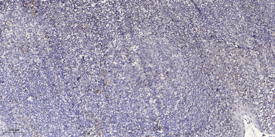
- Immunohistochemical analysis of paraffin-embedded human tonsil. 1, Antibody was diluted at 1:200(4° overnight). 2, Tris-EDTA,pH9.0 was used for antigen retrieval. 3,Secondary antibody was diluted at 1:200(room temperature, 45min).
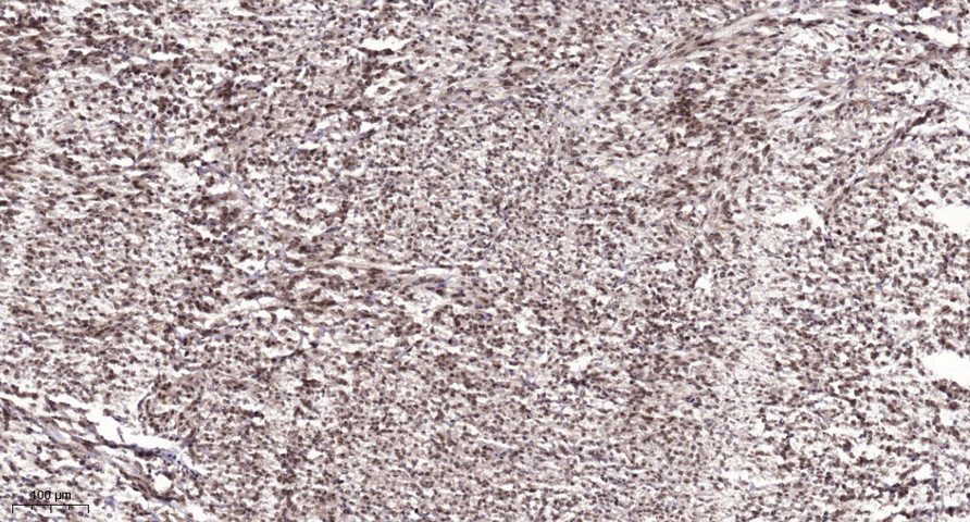
- Immunohistochemical analysis of paraffin-embedded human Colon cancer. 1, Antibody was diluted at 1:200(4° overnight). 2, Tris-EDTA,pH9.0 was used for antigen retrieval. 3,Secondary antibody was diluted at 1:200(room temperature, 45min).


