CD68 Monoclonal Antibody(6F3)
- Catalog No.:YM3050
- Applications:IHC;IF
- Reactivity:Human;Mouse;Rat
- Target:
- CD68
- Fields:
- >>Lysosome
- Gene Name:
- CD68
- Protein Name:
- Macrosialin
- Human Gene Id:
- 968
- Human Swiss Prot No:
- P34810
- Mouse Gene Id:
- 12514
- Mouse Swiss Prot No:
- P31996
- Immunogen:
- Synthetic Peptide of CD68
- Specificity:
- The antibody detects endogenous CD68 proteins.
- Formulation:
- PBS, pH 7.4, containing 0.5%BSA, 0.02% sodium azide as Preservative and 50% Glycerol.
- Source:
- Monoclonal, Mouse
- Dilution:
- IHC 1:200-400, IF 1:50-200
- Purification:
- The antibody was affinity-purified from mouse ascites by affinity-chromatography using specific immunogen.
- Storage Stability:
- -15°C to -25°C/1 year(Do not lower than -25°C)
- Other Name:
- CD68;Macrosialin;Gp110;CD68
- Observed Band(KD):
- 37kD
- Background:
- This gene encodes a 110-kD transmembrane glycoprotein that is highly expressed by human monocytes and tissue macrophages. It is a member of the lysosomal/endosomal-associated membrane glycoprotein (LAMP) family. The protein primarily localizes to lysosomes and endosomes with a smaller fraction circulating to the cell surface. It is a type I integral membrane protein with a heavily glycosylated extracellular domain and binds to tissue- and organ-specific lectins or selectins. The protein is also a member of the scavenger receptor family. Scavenger receptors typically function to clear cellular debris, promote phagocytosis, and mediate the recruitment and activation of macrophages. Alternative splicing results in multiple transcripts encoding different isoforms. [provided by RefSeq, Jul 2008],
- Function:
- function:Could play a role in phagocytic activities of tissue macrophages, both in intracellular lysosomal metabolism and extracellular cell-cell and cell-pathogen interactions. Bind to tissue- and organ-specific lectins or selectins, allowing homing of macrophage subsets to particular sites. Rapid recirculation of CD68 from endosomes, lysosomes to the plasma membrane may allow macrophages to crawl over selectin bearing substrates or other cells.,PTM:N- and O-glycosylated.,similarity:Belongs to the LAMP family.,tissue specificity:Highly expressed by blood monocytes and tissue macrophages. Also expressed in many tumor cell lines which could allow them to attach to selectins on vascular endothelium, facilitating their dissemination to secondary sites.,
- Subcellular Location:
- [Isoform Short]: Cell membrane; Single-pass type I membrane protein.; [Isoform Long]: Endosome membrane; Single-pass type I membrane protein. Lysosome membrane; Single-pass type I membrane protein.
- Expression:
- Highly expressed by blood monocytes and tissue macrophages. Also expressed in lymphocytes, fibroblasts and endothelial cells. Expressed in many tumor cell lines which could allow them to attach to selectins on vascular endothelium, facilitating their dissemination to secondary sites.
Mitophagy-promoting miR-138-5p promoter demethylation inhibits pyroptosis in sepsis-associated acute lung injury INFLAMMATION RESEARCH Liu, Fen, Yang, Ying, Peng, Wei, Zhao, Ning, Chen, Jiaquan, Xu, Zeyao, Cui, Yamei, Qian, Kejian IF Mouse lung tissue
7-Difluoromethoxy-5,4′-dimethoxy-genistein attenuates macrophages apoptosis to promote plaque stability via TIPE2/TLR4 axis in high fat diet-fed ApoE−/− mice. INTERNATIONAL IMMUNOPHARMACOLOGY Int Immunopharmacol. 2021 Jul;96:107477 IHC,IF Mouse 1 : 100 ,1:200 Aortic tissue
Gucciardo, Erika, et al. "An Ex Vivo Tissue Culture Model for Fibrovascular Complications in Proliferative Diabetic Retinopathy." JoVE (Journal of Visualized Experiments) 143 (2019): e59090.
7-Difluoromethoxy-5, 4′-dimethoxy-genistein attenuates macrophages apoptosis to promote plaque stability via TIPE2/TLR4 axis in high fat diet-fed ApoE−/− mice." International Immunopharmacology 96 (2021): 107477.
Long-Chain Acyl Carnitines Aggravate Polystyrene Nanoplastics-Induced Atherosclerosis by Upregulating MARCO. Zhenlie Huang IF Mouse aorta
H3.3B controls aortic dissection progression by regulating vascular smooth muscle cells phenotypic transition and vascular inflammation. Li Wang IHC Mouse aortic tissues
Behind the Indolent Facade: Uncovering the Molecular Features and Malignancy Potential in Lung Minimally Invasive Adenocarcinoma by Single-Cell Transcriptomics Advanced Science Zhenlie Huang IF Human lung adenocarcinoma (LUAD) tissue
- June 19-2018
- WESTERN IMMUNOBLOTTING PROTOCOL
- June 19-2018
- IMMUNOHISTOCHEMISTRY-PARAFFIN PROTOCOL
- June 19-2018
- IMMUNOFLUORESCENCE PROTOCOL
- September 08-2020
- FLOW-CYTOMEYRT-PROTOCOL
- May 20-2022
- Cell-Based ELISA│解您多样本WB检测之困扰
- July 13-2018
- CELL-BASED-ELISA-PROTOCOL-FOR-ACETYL-PROTEIN
- July 13-2018
- CELL-BASED-ELISA-PROTOCOL-FOR-PHOSPHO-PROTEIN
- July 13-2018
- Antibody-FAQs
- Products Images
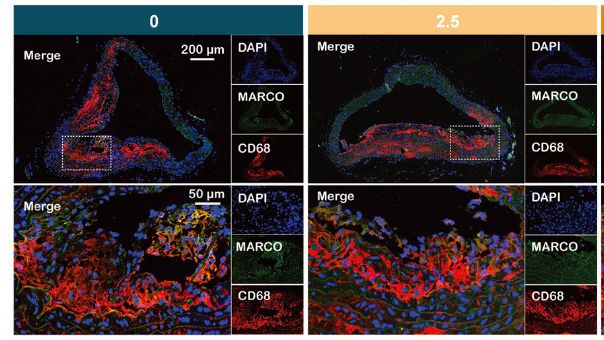
- Long-Chain Acyl Carnitines Aggravate Polystyrene Nanoplastics-Induced Atherosclerosis by Upregulating MARCO. Zhenlie Huang IF Mouse aorta
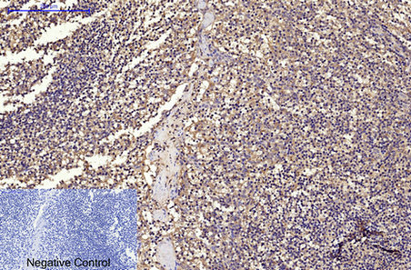
- Immunohistochemical analysis of paraffin-embedded Human-Tonsil tissue. 1,CD68 Monoclonal Antibody(6F3) was diluted at 1:200(4°C,overnight). 2, Sodium citrate pH 6.0 was used for antibody retrieval(>98°C,20min). 3,Secondary antibody was diluted at 1:200(room tempeRature, 30min). Negative control was used by secondary antibody only.
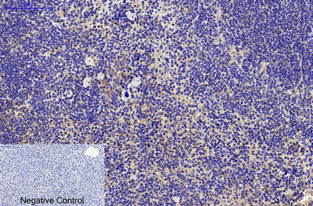
- Immunohistochemical analysis of paraffin-embedded Mouse-liver tissue. 1,CD68 Monoclonal Antibody(6F3) was diluted at 1:200(4°C,overnight). 2, Sodium citrate pH 6.0 was used for antibody retrieval(>98°C,20min). 3,Secondary antibody was diluted at 1:200(room tempeRature, 30min). Negative control was used by secondary antibody only.
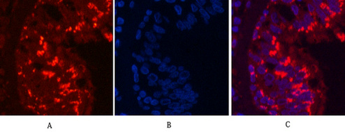
- Immunofluorescence analysis of Human-lung-cancer tissue. 1,CD68 Monoclonal Antibody(6F3)(red) was diluted at 1:200(4°C,overnight). 2, Cy3 labled Secondary antibody was diluted at 1:300(room temperature, 50min).3, Picture B: DAPI(blue) 10min. Picture A:Target. Picture B: DAPI. Picture C: merge of A+B
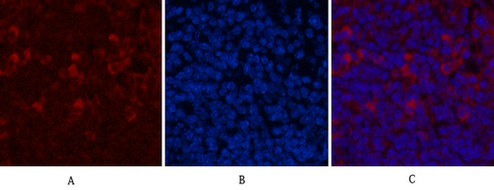
- Immunofluorescence analysis of Mouse-spleen tissue. 1,CD68 Monoclonal Antibody(6F3)(red) was diluted at 1:200(4°C,overnight). 2, Cy3 labled Secondary antibody was diluted at 1:300(room temperature, 50min).3, Picture B: DAPI(blue) 10min. Picture A:Target. Picture B: DAPI. Picture C: merge of A+B
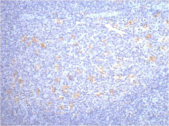
- IHC staining of human tonsil tissue, diluted at 1:200.


