WIF-1 Monoclonal Antibody
- Catalog No.:YM0648
- Applications:WB;IHC;IF;ELISA
- Reactivity:Human
- Target:
- WIF-1
- Fields:
- >>Wnt signaling pathway
- Gene Name:
- WIF1
- Protein Name:
- Wnt inhibitory factor 1
- Human Gene Id:
- 11197
- Human Swiss Prot No:
- Q9Y5W5
- Mouse Swiss Prot No:
- Q9WUA1
- Immunogen:
- Purified recombinant fragment of human WIF-1 expressed in E. Coli.
- Specificity:
- WIF-1 Monoclonal Antibody detects endogenous levels of WIF-1 protein.
- Formulation:
- Liquid in PBS containing 50% glycerol, 0.5% BSA and 0.02% sodium azide.
- Source:
- Monoclonal, Mouse
- Dilution:
- WB 1:500 - 1:2000. IHC 1:200 - 1:1000. IF 1:200 - 1:1000. ELISA: 1:10000. Not yet tested in other applications.
- Purification:
- Affinity purification
- Storage Stability:
- -15°C to -25°C/1 year(Do not lower than -25°C)
- Other Name:
- WIF1;Wnt inhibitory factor 1;WIF-1
- Molecular Weight(Da):
- 42kD
- References:
- 1. BMC Cancer. 2009 Jul 1;9:217.
2. Cancer Res. 2009 Nov 15;69(22):8603-10.
- Background:
- The protein encoded by this gene functions to inhibit WNT proteins, which are extracellular signaling molecules that play a role in embryonic development. This protein contains a WNT inhibitory factor (WIF) domain and five epidermal growth factor (EGF)-like domains, and is thought to be involved in mesoderm segmentation. This gene functions as a tumor suppressor gene, and has been found to be epigenetically silenced in various cancers. [provided by RefSeq, Jun 2010],
- Function:
- function:Binds to WNT proteins and inhibits their activities. May be involved in mesoderm segmentation.,similarity:Contains 1 WIF domain.,similarity:Contains 5 EGF-like domains.,
- Subcellular Location:
- Secreted.
- Expression:
- Brain,
Single-Cell RNA Sequencing Reveals the Cellular Origin and Evolution of Breast Cancer in BRCA1 Mutation CarriersThe Cell Origin of BRCA1-Mutated Breast Cancer. CANCER RESEARCH Cancer Res. 2021 May;81(10):2600-2611 IHC Human normal breast tissues
- June 19-2018
- WESTERN IMMUNOBLOTTING PROTOCOL
- June 19-2018
- IMMUNOHISTOCHEMISTRY-PARAFFIN PROTOCOL
- June 19-2018
- IMMUNOFLUORESCENCE PROTOCOL
- September 08-2020
- FLOW-CYTOMEYRT-PROTOCOL
- May 20-2022
- Cell-Based ELISA│解您多样本WB检测之困扰
- July 13-2018
- CELL-BASED-ELISA-PROTOCOL-FOR-ACETYL-PROTEIN
- July 13-2018
- CELL-BASED-ELISA-PROTOCOL-FOR-PHOSPHO-PROTEIN
- July 13-2018
- Antibody-FAQs
- Products Images
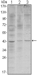
- Western Blot analysis using WIF-1 Monoclonal Antibody against HeLa (1), NIH/3T3 (2) and NTERA-2 (3) cell lysate.
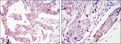
- Immunohistochemistry analysis of paraffin-embedded ovary tumour tissues (left) and lung cancer (right) with DAB staining using WIF-1 Monoclonal Antibody.
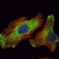
- Immunofluorescence analysis of Hela cells using WIF-1 Monoclonal Antibody (green). Red: Actin filaments have been labeled with Alexa Fluor-555 phalloidin.


