Calcyclin Monoclonal Antibody
- Catalog No.:YM0088
- Applications:WB;IHC;IF;ELISA
- Reactivity:Human
- Target:
- Calcyclin
- Fields:
- >>Wnt signaling pathway
- Gene Name:
- CACYBP
- Protein Name:
- Calcyclin-binding protein
- Human Gene Id:
- 27101
- Human Swiss Prot No:
- Q9HB71
- Mouse Swiss Prot No:
- Q9CXW3
- Immunogen:
- Purified recombinant fragment of Calcyclin expressed in E. Coli.
- Specificity:
- Calcyclin Monoclonal Antibody detects endogenous levels of Calcyclin protein.
- Formulation:
- Liquid in PBS containing 50% glycerol, 0.5% BSA and 0.02% sodium azide.
- Source:
- Monoclonal, Mouse
- Dilution:
- WB 1:500 - 1:2000. IHC 1:200 - 1:1000. ELISA: 1:10000.. IF 1:50-200
- Purification:
- Affinity purification
- Concentration:
- 1 mg/ml
- Storage Stability:
- -15°C to -25°C/1 year(Do not lower than -25°C)
- Other Name:
- CACYBP;S100A6BP;SIP;PNAS-107;Calcyclin-binding protein;CacyBP;hCacyBP;S100A6-binding protein;Siah-interacting protein
- Molecular Weight(Da):
- 26kD
- References:
- 1. Bean C.et.al J Mol Biol. 2005 Jun 3;349(2):349-66.
2. HWang R.et.al J Biol Chem. 2004 May 14;279(20):21239-47.
- Background:
- The protein encoded by this gene is a calcyclin binding protein. It may be involved in calcium-dependent ubiquitination and subsequent proteosomal degradation of target proteins. It probably serves as a molecular bridge in ubiquitin E3 complexes and participates in the ubiquitin-mediated degradation of beta-catenin. Two alternatively spliced transcript variants encoding different isoforms have been found for this gene. [provided by RefSeq, Jul 2008],
- Function:
- function:May be involved in calcium-dependent ubiquitination and subsequent proteosomal degradation of target proteins. Probably serves as a molecular bridge in ubiquitin E3 complexes. Participates in the ubiquitin-mediated degradation of beta-catenin (CTNNB1).,PTM:Phosphorylated on serine residues. Phosphorylated upon induction by RA or at high calcium concentrations.,similarity:Contains 1 CS domain.,similarity:Contains 1 SGS domain.,subcellular location:Cytoplasmic at low calcium concentrations. In neuroblastoma cells, after a retinoic acid (RA) induction and calcium increase, it localizes in both the nucleus and cytoplasm. The nuclear fraction may be phosphorylated.,subunit:Interacts with protein of the S100 family S100A1, S100A6, S100B, S100P and S100A12 at physiological calcium concentrations (By similarity). Component of some large E3 complex at least composed of UBE2D1, SIAH1, CAC
- Subcellular Location:
- Nucleus . Cytoplasm . Cytoplasmic at low calcium concentrations. In neuroblastoma cells, after a retinoic acid (RA) induction and calcium increase, it localizes in both the nucleus and cytoplasm. The nuclear fraction may be phosphorylated.
- Expression:
- Brain,Colon carcinoma,Hepatoma,Liver,Promyelocytic leukemia,Skeletal muscle
- June 19-2018
- WESTERN IMMUNOBLOTTING PROTOCOL
- June 19-2018
- IMMUNOHISTOCHEMISTRY-PARAFFIN PROTOCOL
- June 19-2018
- IMMUNOFLUORESCENCE PROTOCOL
- September 08-2020
- FLOW-CYTOMEYRT-PROTOCOL
- May 20-2022
- Cell-Based ELISA│解您多样本WB检测之困扰
- July 13-2018
- CELL-BASED-ELISA-PROTOCOL-FOR-ACETYL-PROTEIN
- July 13-2018
- CELL-BASED-ELISA-PROTOCOL-FOR-PHOSPHO-PROTEIN
- July 13-2018
- Antibody-FAQs
- Products Images
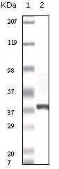
- Western Blot analysis using Calcyclin Monoclonal Antibody against truncated calcyclin recombinant protein.
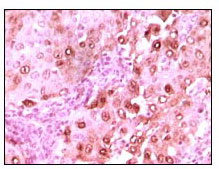
- Immunohistochemistry analysis of paraffin-embedded human brain glioma tissue showing nuclear localization with DAB staining using Calcyclin Monoclonal Antibody.


