AIF-M1 Monoclonal Antibody
- Catalog No.:YM0017
- Applications:WB;IHC;IF;FCM;ELISA
- Reactivity:Human;Mouse;Rat;Monkey
- Target:
- AIF-M1
- Fields:
- >>Apoptosis;>>Necroptosis
- Gene Name:
- AIFM1
- Protein Name:
- Apoptosis-inducing factor 1 mitochondrial
- Human Gene Id:
- 9131
- Human Swiss Prot No:
- O95831
- Mouse Gene Id:
- 26926
- Mouse Swiss Prot No:
- Q9Z0X1
- Rat Gene Id:
- 83533
- Rat Swiss Prot No:
- Q9JM53
- Immunogen:
- Purified recombinant fragment of human AIF-M1 expressed in E. Coli.
- Specificity:
- AIF-M1 Monoclonal Antibody detects endogenous levels of AIF-M1 protein.
- Formulation:
- Liquid in PBS containing 50% glycerol, 0.5% BSA and 0.02% sodium azide.
- Source:
- Monoclonal, Mouse
- Dilution:
- WB 1:500 - 1:2000. IHC 1:200 - 1:1000. IF 1:200 - 1:1000. Flow cytometry: 1:200 - 1:400. ELISA: 1:10000. Not yet tested in other applications.
- Purification:
- Affinity purification
- Storage Stability:
- -15°C to -25°C/1 year(Do not lower than -25°C)
- Other Name:
- AIFM1;AIF;PDCD8;Apoptosis-inducing factor 1; mitochondrial;Programmed cell death protein 8
- Molecular Weight(Da):
- 67kD
- References:
- 1. Apoptosis. 2009 Jun;14(6):796-808.
2. PLoS One. 2009;4(2):e4394. Epub 2009 Feb 6.
- Background:
- This gene encodes a flavoprotein essential for nuclear disassembly in apoptotic cells, and it is found in the mitochondrial intermembrane space in healthy cells. Induction of apoptosis results in the translocation of this protein to the nucleus where it affects chromosome condensation and fragmentation. In addition, this gene product induces mitochondria to release the apoptogenic proteins cytochrome c and caspase-9. Mutations in this gene cause combined oxidative phosphorylation deficiency 6 (COXPD6), a severe mitochondrial encephalomyopathy, as well as Cowchock syndrome, also known as X-linked recessive Charcot-Marie-Tooth disease-4 (CMTX-4), a disorder resulting in neuropathy, and axonal and motor-sensory defects with deafness and mental retardation. Alternative splicing results in multiple transcript variants. A related pseudogene has been identified on chromosome
- Function:
- catalytic activity:2 glutathione + protein-disulfide = glutathione disulfide + protein-dithiol.,cofactor:FAD.,function:Possesses significant protein thiol-disulfide oxidase activity.,function:Probable oxidoreductase that acts as a caspase-independent mitochondrial effector of apoptotic cell death. Extramitochondrial AIF induces nuclear chromatin condensation and large scale DNA fragmentation (in vitro). Binds to DNA in a sequence-independent manner.,similarity:Belongs to the FAD-dependent oxidoreductase family.,similarity:Contains 1 thioredoxin domain.,subcellular location:Translocated to the nucleus upon induction of apoptosis.,subunit:Interacts with XIAP.,tissue specificity:Widely expressed.,
- Subcellular Location:
- Mitochondrion intermembrane space . Mitochondrion inner membrane. Cytoplasm . Nucleus . Cytoplasm, perinuclear region . Proteolytic cleavage during or just after translocation into the mitochondrial intermembrane space (IMS) results in the formation of an inner-membrane-anchored mature form (AIFmit). During apoptosis, further proteolytic processing leads to a mature form, which is confined to the mitochondrial IMS in a soluble form (AIFsol). AIFsol is released to the cytoplasm in response to specific death signals, and translocated to the nucleus, where it induces nuclear apoptosis (PubMed:15775970). Colocalizes with EIF3G in the nucleus and perinuclear region (PubMed:17094969). .; [Isoform 3]: Mitochondrion intermembrane space . Mitochondrion inner membrane . Has a stronger membrane ancho
- Expression:
- Expressed in all tested tissues (PubMed:16644725). Detected in muscle and skin fibroblasts (at protein level) (PubMed:23217327). Expressed in osteoblasts (at protein level) (PubMed:28842795). ; [Isoform 3]: Brain specific. ; [Isoform 4]: Expressed in all tested tissues except brain. ; [Isoform 5]: Isoform 5 is frequently down-regulated in human cancers.
- June 19-2018
- WESTERN IMMUNOBLOTTING PROTOCOL
- June 19-2018
- IMMUNOHISTOCHEMISTRY-PARAFFIN PROTOCOL
- June 19-2018
- IMMUNOFLUORESCENCE PROTOCOL
- September 08-2020
- FLOW-CYTOMEYRT-PROTOCOL
- May 20-2022
- Cell-Based ELISA│解您多样本WB检测之困扰
- July 13-2018
- CELL-BASED-ELISA-PROTOCOL-FOR-ACETYL-PROTEIN
- July 13-2018
- CELL-BASED-ELISA-PROTOCOL-FOR-PHOSPHO-PROTEIN
- July 13-2018
- Antibody-FAQs
- Products Images
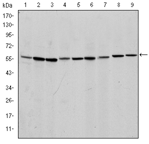
- Western Blot analysis using AIF-M1 Monoclonal Antibody against NIH/3T3 (1), Jurkat (2), HeLa (3), HepG2 (4), MOLT4 (5), C6 (6), RAJI (7), Cos7 (8) and PC-12 (9) cell lysate.
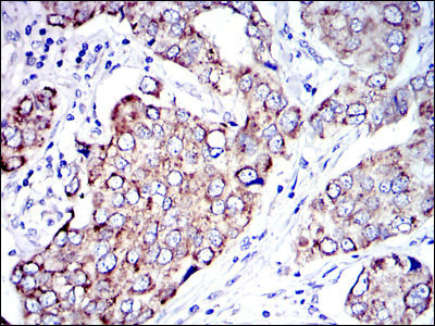
- Immunohistochemistry analysis of paraffin-embedded human breast cancer tissues with DAB staining using AIF-M1 Monoclonal Antibody.
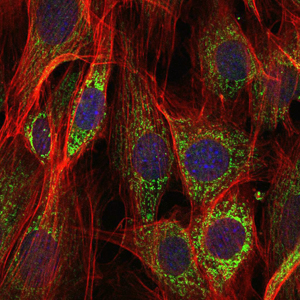
- Immunofluorescence analysis of NIH/3T3 cells using AIF-M1 Monoclonal Antibody (green). Blue: DRAQ5 fluorescent DNA dye. Red: Actin filaments have been labeled with Alexa Fluor-555 phalloidin.
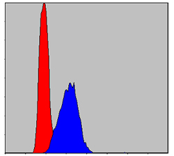
- Flow cytometric analysis of HepG2 cells using AIF-M1 Monoclonal Antibody (blue) and negative control (red).


