- 靶点:
- OPG
- 简介:
- >>Cytokine-cytokine receptor interaction;>>Osteoclast differentiation
- 基因名称:
- TNFRSF11B
- 蛋白名称:
- Tumor necrosis factor receptor superfamily member 11B
- Human Gene Id:
- 4982
- Human Swiss Prot No:
- O00300
- Mouse Gene Id:
- 18383
- Mouse Swiss Prot No:
- O08712
- 免疫原:
- The antiserum was produced against synthesized peptide derived from human TR11B . AA range:10-59
- 特异性:
- OPG Polyclonal Antibody detects endogenous levels of OPG protein.
- 组成:
- Liquid in PBS containing 50% glycerol, 0.5% BSA and 0.02% sodium azide.
- 来源:
- Polyclonal, Rabbit,IgG
- 稀释:
- WB 1:500 - 1:2000. IHC: 1:100-300 ELISA: 1:20000. IF 1:100-300 Not yet tested in other applications.
- 纯化工艺:
- The antibody was affinity-purified from rabbit antiserum by affinity-chromatography using epitope-specific immunogen.
- 浓度:
- 1 mg/ml
- 储存:
- -15°C to -25°C/1 year(Do not lower than -25°C)
- 其他名称:
- TNFRSF11B;OCIF;OPG;Tumor necrosis factor receptor superfamily member 11B;Osteoclastogenesis inhibitory factor;Osteoprotegerin
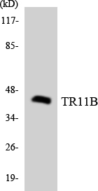
- 实测条带:
- 55kD
- 背景:
- The protein encoded by this gene is a member of the TNF-receptor superfamily. This protein is an osteoblast-secreted decoy receptor that functions as a negative regulator of bone resorption. This protein specifically binds to its ligand, osteoprotegerin ligand, both of which are key extracellular regulators of osteoclast development. Studies of the mouse counterpart also suggest that this protein and its ligand play a role in lymph-node organogenesis and vascular calcification. Alternatively spliced transcript variants of this gene have been reported, but their full length nature has not been determined. [provided by RefSeq, Jul 2008],
- 功能:
- disease:Defects in TNFRSF11B are the cause of juvenile Paget disease (JPD) [MIM:239000]; also called hyperostosis corticalis deformans juvenilis or hereditary hyperphosphatasia or chronic congenital idiopathic hyperphosphatasia. JPD is a rare autosomal recessive osteopathy that presents in infancy or early childhood. The disorder is characterized by rapidly remodeling woven bone, osteopenia, debilitating fractures, and deformities due to a markedly accelerated rate of bone remodeling throughout the skeleton. Approximately 40 cases of JPD have been reported worldwide. Unless it is treated with drugs that block osteoclast-mediated skeletal resorption, the disease can be fatal.,function:Acts as decoy receptor for RANKL and thereby neutralizes its function in osteoclastogenesis. Inhibits the activation of osteoclasts and promotes osteoclast apoptosis in vitro. Bone homeostasis seems to depen
- 细胞定位:
- Secreted.
- 组织表达:
- Highly expressed in adult lung, heart, kidney, liver, spleen, thymus, prostate, ovary, small intestine, thyroid, lymph node, trachea, adrenal gland, testis, and bone marrow. Detected at very low levels in brain, placenta and skeletal muscle. Highly expressed in fetal kidney, liver and lung.
O-Linked N-Acetylglucosamine Transferase Regulates Bone Homeostasis Through Alkaline Phosphatase Pathway in Diabetic Periodontitis MOLECULAR BIOTECHNOLOGY Luo Wei IHC,WB Human,Mouse 1:100,1:1000 gingival tissue Human periodontal ligament cells (hPDLCs)
货号:YT3466
- June 19-2018
- WESTERN IMMUNOBLOTTING PROTOCOL
- June 19-2018
- IMMUNOHISTOCHEMISTRY-PARAFFIN PROTOCOL
- June 19-2018
- IMMUNOFLUORESCENCE PROTOCOL
- September 08-2020
- FLOW-CYTOMEYRT-PROTOCOL
- May 20-2022
- Cell-Based ELISA│解您多样本WB检测之困扰
- July 13-2018
- CELL-BASED-ELISA-PROTOCOL-FOR-ACETYL-PROTEIN
- July 13-2018
- CELL-BASED-ELISA-PROTOCOL-FOR-PHOSPHO-PROTEIN
- July 13-2018
- Antibody-FAQs
- 产品图片
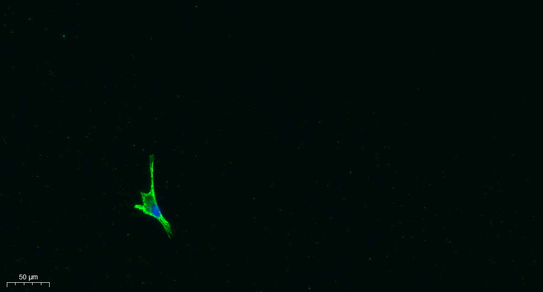
- Immunofluorescence analysis of A549. 1,primary Antibody was diluted at 1:200(4°C overnight). 2, Goat Anti Rabbit IgG (H&L) - Alexa Fluor 488 Secondary antibody was diluted at 1:1000(room temperature, 50min).3, Picture B: DAPI(blue) 10min.
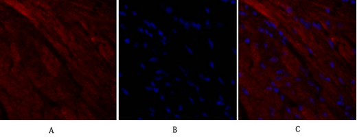
- Immunofluorescence analysis of mouse-heart tissue. 1,OPG Polyclonal Antibody(red) was diluted at 1:200(4°C,overnight). 2, Cy3 labled Secondary antibody was diluted at 1:300(room temperature, 50min).3, Picture B: DAPI(blue) 10min. Picture A:Target. Picture B: DAPI. Picture C: merge of A+B
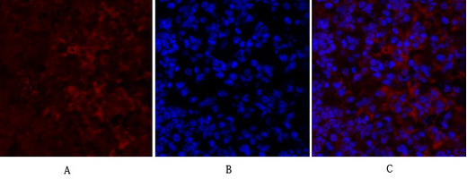
- Immunofluorescence analysis of mouse-lung tissue. 1,OPG Polyclonal Antibody(red) was diluted at 1:200(4°C,overnight). 2, Cy3 labled Secondary antibody was diluted at 1:300(room temperature, 50min).3, Picture B: DAPI(blue) 10min. Picture A:Target. Picture B: DAPI. Picture C: merge of A+B
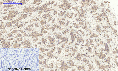
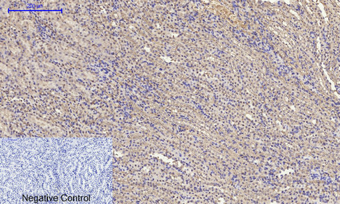
- Immunohistochemical analysis of paraffin-embedded Human-liver-cancer tissue. 1,OPG Polyclonal Antibody was diluted at 1:200(4°C,overnight). 2, Sodium citrate pH 6.0 was used for antibody retrieval(>98°C,20min). 3,Secondary antibody was diluted at 1:200(room tempeRature, 30min). Negative control was used by secondary antibody only.
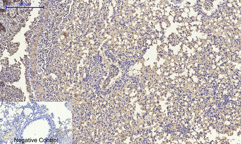
- Immunohistochemical analysis of paraffin-embedded Mouse-lung tissue. 1,OPG Polyclonal Antibody was diluted at 1:200(4°C,overnight). 2, Sodium citrate pH 6.0 was used for antibody retrieval(>98°C,20min). 3,Secondary antibody was diluted at 1:200(room tempeRature, 30min). Negative control was used by secondary antibody only.
- Immunohistochemical analysis of paraffin-embedded Mouse-kidney tissue. 1,OPG Polyclonal Antibody was diluted at 1:200(4°C,overnight). 2, Sodium citrate pH 6.0 was used for antibody retrieval(>98°C,20min). 3,Secondary antibody was diluted at 1:200(room tempeRature, 30min). Negative control was used by secondary antibody only.
- Western Blot analysis of various cells using OPG Polyclonal Antibody diluted at 1:1000
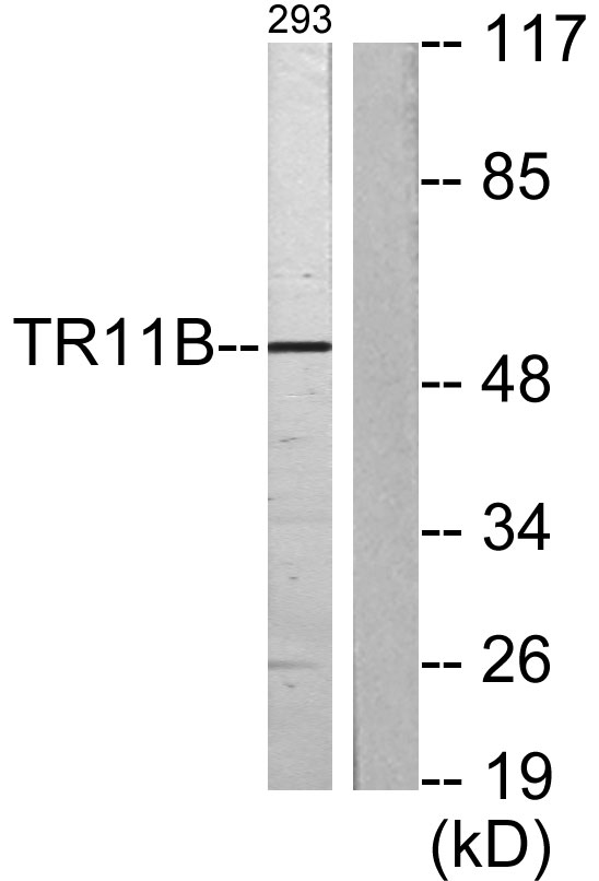
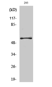
- Western blot analysis of lysates from 293 cells, using TR11B Antibody . The lane on the right is blocked with the synthesized peptide.
- Western blot analysis of the lysates from HeLa cells using TR11B antibody.


