- 靶点:
- CD31
- 简介:
- >>Cell adhesion molecules;>>Leukocyte transendothelial migration;>>Malaria;>>Fluid shear stress and atherosclerosis
- 基因名称:
- PECAM1
- 蛋白名称:
- Platelet endothelial cell adhesion molecule (PECAM-1) (EndoCAM) (GPIIA') (PECA1) (CD antigen CD31)
- Human Gene Id:
- 5175
- Human Swiss Prot No:
- P16284
- 免疫原:
- Synthesized peptide derived from human CD31 AA range: 100-200
- 特异性:
- This antibody detects endogenous levels of human CD31. Heat-induced epitope retrieval (HIER) Citrate buffer of pH6.0 was highly recommended as antigen repair method in paraffin section.
- 组成:
- Liquid in PBS containing 50% glycerol, 0.5% BSA and 0.02% sodium azide.
- 来源:
- Mouse, Monoclonal/IgG1, Kappa
- 稀释:
- WB 1:200 - 1:500, IHC 1:20-50, IF 1:200 - 1:1000, ELISA: 1:10000
- 纯化工艺:
- The antibody was affinity-purified from mouse ascites by affinity-chromatography using specific immunogen.
- 储存:
- -15°C to -25°C/1 year(Do not lower than -25°C)
- 分子量:
- 83kD
- 背景:
- The protein encoded by this gene is found on the surface of platelets, monocytes, neutrophils, and some types of T-cells, and makes up a large portion of endothelial cell intercellular junctions. The encoded protein is a member of the immunoglobulin superfamily and is likely involved in leukocyte migration, angiogenesis, and integrin activation. [provided by RefSeq, May 2010],
- 功能:
- function:This protein is a cell adhesion molecule expressed on platelets and at endothelial cell intercellular junctions.,online information:CD31 entry,online information:PECAM-1,online information:The Singapore human mutation and polymorphism database,PTM:Phosphorylated on Ser and Tyr residues after cellular activation.,similarity:Contains 6 Ig-like C2-type (immunoglobulin-like) domains.,tissue specificity:Long isoform predominates all tissues examined, isoform Delta12 was detected only in trachea and isoform Delta14-15 only in lung, isoform Delta14 was detected in all tissues examined with the strongest expression in heart.,
- 细胞定位:
- Cell membrane ; Single-pass type I membrane protein . Cell surface expression on neutrophils is down-regulated upon fMLP or CXCL8/IL8-mediated stimulation. .; [Isoform Long]: Cell membrane ; Single-pass type I membrane protein . Membrane raft . Cell junction . Localizes to the lateral border recycling compartment (LBRC) and recycles from the LBRC to the junction in resting endothelial cells. .; [Isoform Delta15]: Cell junction . Localizes to the lateral border recycling compartment (LBRC) and recycles from the LBRC to the junction in resting endothelial cells.
- 组织表达:
- Expressed on platelets and leukocytes and is primarily concentrated at the borders between endothelial cells (PubMed:18388311, PubMed:21464369). Expressed in human umbilical vein endothelial cells (HUVECs) (at protein level) (PubMed:19342684, PubMed:17580308). Expressed on neutrophils (at protein level) (PubMed:17580308). Isoform Long predominates in all tissues examined (PubMed:12433657). Isoform Delta12 is detected only in trachea (PubMed:12433657). Isoform Delta14-15 is only detected in lung (PubMed:12433657). Isoform Delta14 is detected in all tissues examined with the strongest expression in heart (PubMed:12433657). Isoform Delta15 is expressed in brain, testis, ovary, cell surface of platelets, human umbilical vein endothelial cells (HUVECs), Jurkat T-cell leukemia, human erythroleuk
Systemic administration of mesenchymal stem cells loaded with a novel oncolytic adenovirus carrying IL-24/endostatin enhances glioma therapy. CANCER LETTERS Cancer Lett. 2021 Jul;509:26 IHC Mouse 1 : 200 hUCB-MSCs-Xenograft
货号:YM6115
Direct-writing Process and in vivo Evaluation of Prevascularized Composite Constructs for Muscle Tissue Engineering Application. Journal of Bionic Engineering J Bionic Eng. 2020 May;17(3):457-468 IHC Human Muscle tissue
货号:YM6115
Different stimuli induce endothelial dysfunction and promote atherosclerosis through the Piezo1/YAP signaling axis. ARCHIVES OF BIOCHEMISTRY AND BIOPHYSICS Xiangyu Zhou IF Mouse aortic arch
货号:YM6115
- June 19-2018
- WESTERN IMMUNOBLOTTING PROTOCOL
- June 19-2018
- IMMUNOHISTOCHEMISTRY-PARAFFIN PROTOCOL
- June 19-2018
- IMMUNOFLUORESCENCE PROTOCOL
- September 08-2020
- FLOW-CYTOMEYRT-PROTOCOL
- May 20-2022
- Cell-Based ELISA│解您多样本WB检测之困扰
- July 13-2018
- CELL-BASED-ELISA-PROTOCOL-FOR-ACETYL-PROTEIN
- July 13-2018
- CELL-BASED-ELISA-PROTOCOL-FOR-PHOSPHO-PROTEIN
- July 13-2018
- Antibody-FAQs
- 产品图片
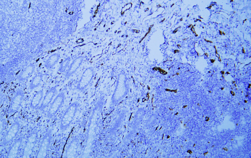
- Human appendix tissue was stained with Anti-CD31 (ABT-CD31) Antibody

- Human Hemangioendothelioma tissue was stained with Anti-CD31 (ABT-CD31) Antibody
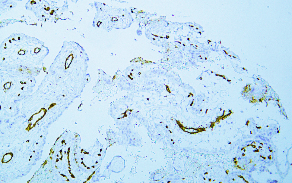
- Human placenta tissue was stained with Anti-CD31 (ABT-CD31) Antibody
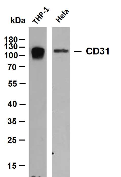
- Various whole cell lysates were separated by 10% SDS-PAGE, and the membrane was blotted with anti-CD31 (ABT-CD31)antibody. The HRP-conjugated Goat anti-Mouse IgG(H + L) antibody was used to detect the antibody. Lane 1: THP-1 Lane 2: Hela Predicted band size: 83kDa Observed band size: 130kDa
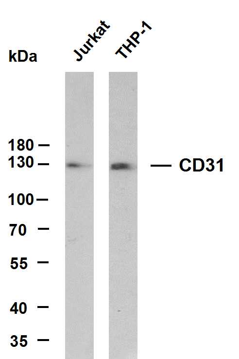
- Various whole cell lysates were separated by 8% SDS-PAGE, and the membrane was blotted with anti-CD31(ABT-CD31) antibody. The HRP-conjugated Goat anti-Mouse IgG(H + L) antibody was used to detect the antibody. Lane 1: Jurkat Lane 2: THP-1 Predicted band size: 83kDa Observed band size: 130kDa
.jpg)
- Immunohistochemical analysis of paraffin-embedded Hemangioendothelioma. 1, Antibody was diluted at 1:200(4° overnight). 2, Citric acid ,pH6.0 was used for antigen retrieval. 3,Secondary antibody was diluted at 1:200(room temperature, 30min).
.jpg)
- Immunohistochemical analysis of paraffin-embedded Hemangioendothelioma. 1, Antibody was diluted at 1:200(4° overnight). 2, Citric acid ,pH6.0 was used for antigen retrieval. 3,Secondary antibody was diluted at 1:200(room temperature, 30min).
.jpg)
- Immunohistochemical analysis of paraffin-embedded Placenta. 1, Antibody was diluted at 1:200(4° overnight). 2, Citric acid ,pH6.0 was used for antigen retrieval. 3,Secondary antibody was diluted at 1:200(room temperature, 30min).
.jpg)
- Immunohistochemical analysis of paraffin-embedded Placenta. 1, Antibody was diluted at 1:200(4° overnight). 2, Citric acid ,pH6.0 was used for antigen retrieval. 3,Secondary antibody was diluted at 1:200(room temperature, 30min).
.jpg)
- Immunohistochemical analysis of paraffin-embedded Placenta. 1, Antibody was diluted at 1:200(4° overnight). 2, Citric acid ,pH6.0 was used for antigen retrieval. 3,Secondary antibody was diluted at 1:200(room temperature, 30min).
.jpg)
- Immunohistochemical analysis of paraffin-embedded Tonsil. 1, Antibody was diluted at 1:200(4° overnight). 2, Citric acid ,pH6.0 was used for antigen retrieval. 3,Secondary antibody was diluted at 1:200(room temperature, 30min).
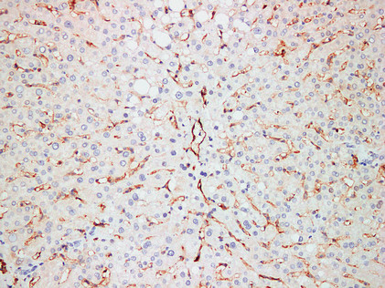
- Immunohistochemical analysis of paraffin-embedded Liver. 1, Antibody was diluted at 1:200(4° overnight). 2, Citric acid ,pH6.0 was used for antigen retrieval. 3,Secondary antibody was diluted at 1:200(room temperature, 30min).
.jpg)
- Immunohistochemical analysis of paraffin-embedded Tonsil. 1, Antibody was diluted at 1:200(4° overnight). 2, Citric acid ,pH6.0 was used for antigen retrieval. 3,Secondary antibody was diluted at 1:200(room temperature, 30min).
_wb.jpg)
- Western blot analysis of CD31Antibody at 1:1000 dilution.


