- 靶点:
- CD146(PT0278)
- 基因名称:
- MCAM MUC18
- 蛋白名称:
- CD146
- Human Gene Id:
- 4162
- Human Swiss Prot No:
- P43121
- 免疫原:
- Synthesized peptide derived from human CD146 AA range: 500-600
- 特异性:
- This antibody detects endogenous levels of human CD146
- 组成:
- Liquid in PBS containing 50% glycerol, 0.5% BSA and 0.02% sodium azide.
- 来源:
- Mouse, Monoclonal/IgG1, Kappa
- 稀释:
- IHC 1:100-300, WB 1:500-1000,ELISA 1:5000-20000
- 纯化工艺:
- The antibody was affinity-purified from mouse ascites by affinity-chromatography using specific immunogen.
- 储存:
- -15°C to -25°C/1 year(Do not lower than -25°C)
- 其他名称:
- Cell surface glycoprotein MUC18 (Cell surface glycoprotein P1H12;Melanoma cell adhesion molecule;Melanoma-associated antigen A32;Melanoma-associated antigen MUC18;S-endo 1 endothelial-associated antigen;CD antigen CD146)
- 分子量:
- 72kD
- 背景:
- function:Plays a role in cell adhesion, and in cohesion of the endothelial monolayer at intercellular junctions in vascular tissue. Its expression may allow melanoma cells to interact with cellular elements of the vascular system, thereby enhancing hematogeneous tumor spread. Could be an adhesion molecule active in neural crest cells during embryonic development. Acts as surface receptor that triggers tyrosine phosphorylation of FYN and PTK2, and a transient increase in the intracellular calcium concentration.,similarity:Contains 2 Ig-like V-type (immunoglobulin-like) domains.,similarity:Contains 3 Ig-like C2-type (immunoglobulin-like) domains.,tissue specificity:Detected in endothelial cells in vascular tissue throughout the body. May appear at the surface of neural crest cells during their embryonic migration. Appears to be limited to vascular smooth muscle in normal adult tissues. Associated with tumor progression and the development of metastasis in human malignant melanoma. Expressed most strongly on metastatic lesions and advanced primary tumors and is only rarely detected in benign melanocytic nevi and thin primary melanomas with a low probability of metastasis.,
- 功能:
- function:Plays a role in cell adhesion, and in cohesion of the endothelial monolayer at intercellular junctions in vascular tissue. Its expression may allow melanoma cells to interact with cellular elements of the vascular system, thereby enhancing hematogeneous tumor spread. Could be an adhesion molecule active in neural crest cells during embryonic development. Acts as surface receptor that triggers tyrosine phosphorylation of FYN and PTK2, and a transient increase in the intracellular calcium concentration.,similarity:Contains 2 Ig-like V-type (immunoglobulin-like) domains.,similarity:Contains 3 Ig-like C2-type (immunoglobulin-like) domains.,tissue specificity:Detected in endothelial cells in vascular tissue throughout the body. May appear at the surface of neural crest cells during their embryonic migration. Appears to be limited to vascular smooth muscle in normal adult tissues. Ass
- 细胞定位:
- Membrane; Single-pass type I membrane protein.
- 组织表达:
- Detected in endothelial cells in vascular tissue throughout the body. May appear at the surface of neural crest cells during their embryonic migration. Appears to be limited to vascular smooth muscle in normal adult tissues. Associated with tumor progression and the development of metastasis in human malignant melanoma. Expressed most strongly on metastatic lesions and advanced primary tumors and is only rarely detected in benign melanocytic nevi and thin primary melanomas with a low probability of metastasis.
A static magnetic field enhances the repair of osteoarthritic cartilage by promoting the migration of stem cells and chondrogenesis Journal of Orthopaedic Translation Ping Zhang IF Mouse knee joint
货号:YM4036
- June 19-2018
- WESTERN IMMUNOBLOTTING PROTOCOL
- June 19-2018
- IMMUNOHISTOCHEMISTRY-PARAFFIN PROTOCOL
- June 19-2018
- IMMUNOFLUORESCENCE PROTOCOL
- September 08-2020
- FLOW-CYTOMEYRT-PROTOCOL
- May 20-2022
- Cell-Based ELISA│解您多样本WB检测之困扰
- July 13-2018
- CELL-BASED-ELISA-PROTOCOL-FOR-ACETYL-PROTEIN
- July 13-2018
- CELL-BASED-ELISA-PROTOCOL-FOR-PHOSPHO-PROTEIN
- July 13-2018
- Antibody-FAQs
- 产品图片
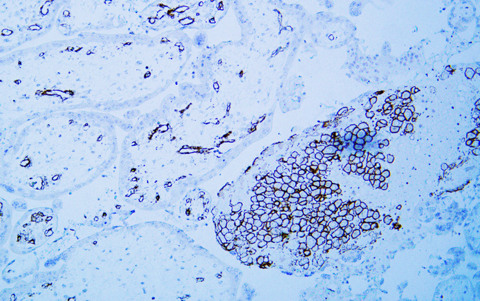
- Human placenta tissue was stained with Anti-CD146 (PT0278) Antibody
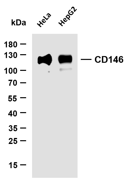
- Various whole cell lysates were separated by 15% SDS-PAGE, and the membrane was blotted with anti-CD146 (PT0278) antibody. The HRP-conjugated Goat anti-Mouse IgG(H + L) antibody was used to detect the antibody. Lane 1: Hela Lane 2: HepG2 Predicted band size: 110kDa Observed band size: 110kDa


